
Structural and Functional Assessments (SFA) Core
Director: Andy Fischer, PhD
Co-director: Colleen Cebulla, MD, PhD
Lab manager: Tyler Heisler-Taylor, PhD
ERG: Julie Racine, PhD
Email: vsrcpmanagement@buckeyemail.osu.edu
Please cite P30EY032857 in all publications that make use of the Ohio State University Vision Science Research Core Program (OSU-VSRCP).
“Services are performed at the OSU Vision Sciences Research Core Program under P30EY032857.”
Services include:
- Technician time & Assisted equipment use ($75/hr)
- Animal handling, husbandry, or other expertise ($40/hr)
- Equipment or procedure training (Free!)
Please reach out or schedule a consultation through eRAMP!
Equipment includes:
-
Diagnosys Celeris ERG System
-
Diagnosys E3 ERG System
-
iCare Tonovet Plus
-
HM 550 Cryostat
-
Leica R2200 SDOIS
-
Micron IV
-
Nikon AXR Confocal
-
Operating Microscope
-
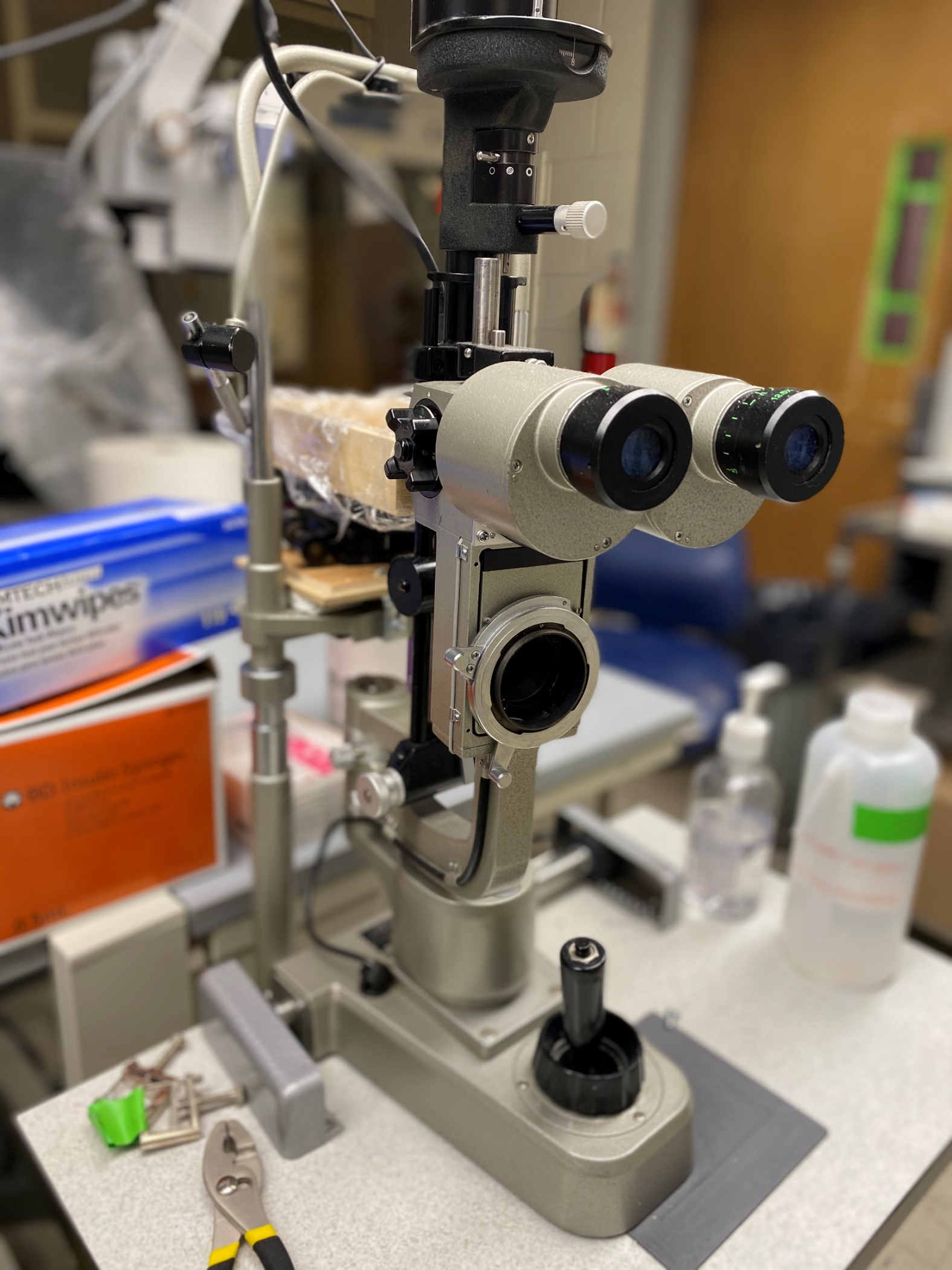
Slit Lamp
-
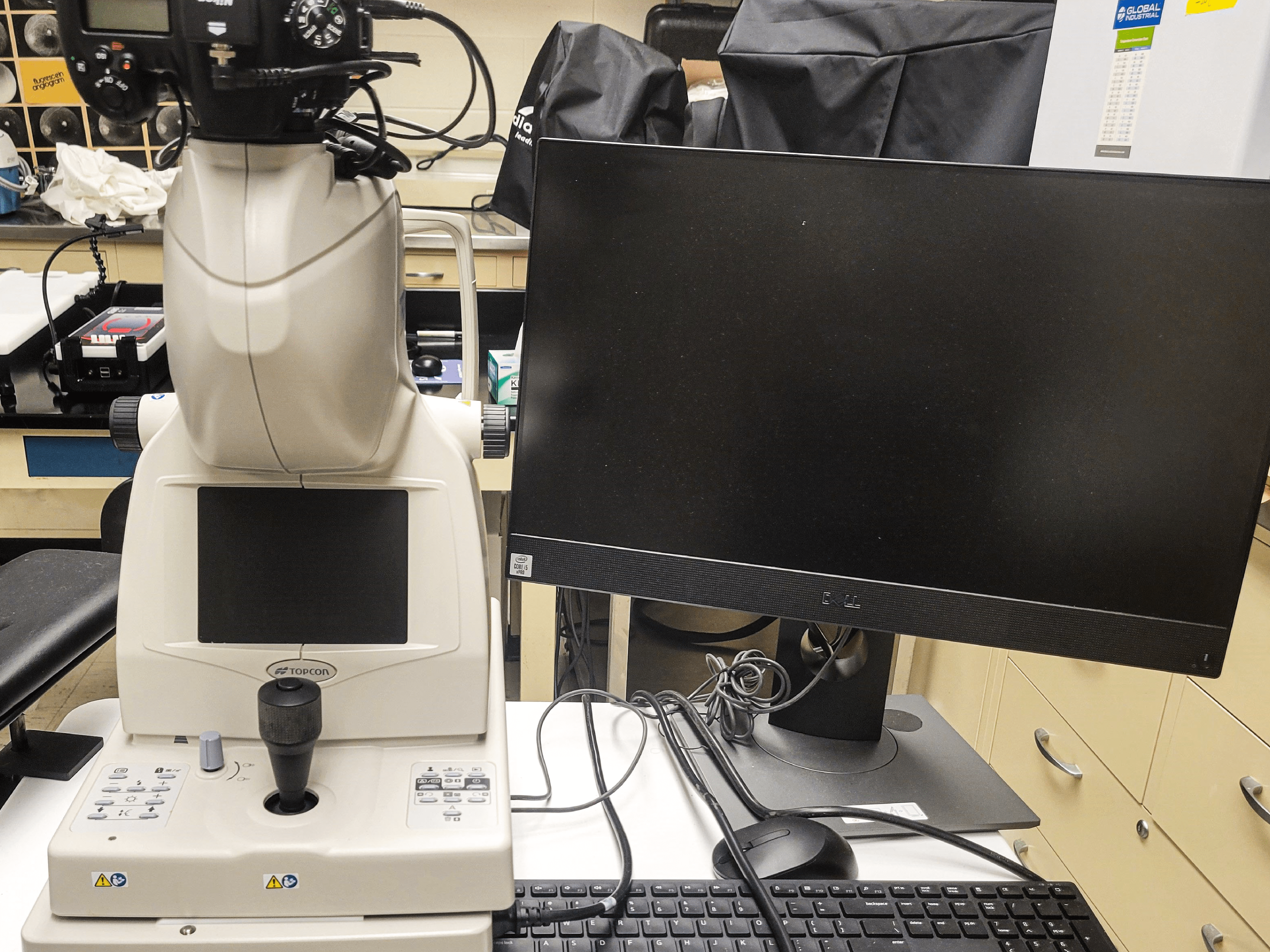
Topcon TRC-NW8F Fundus Camera
-
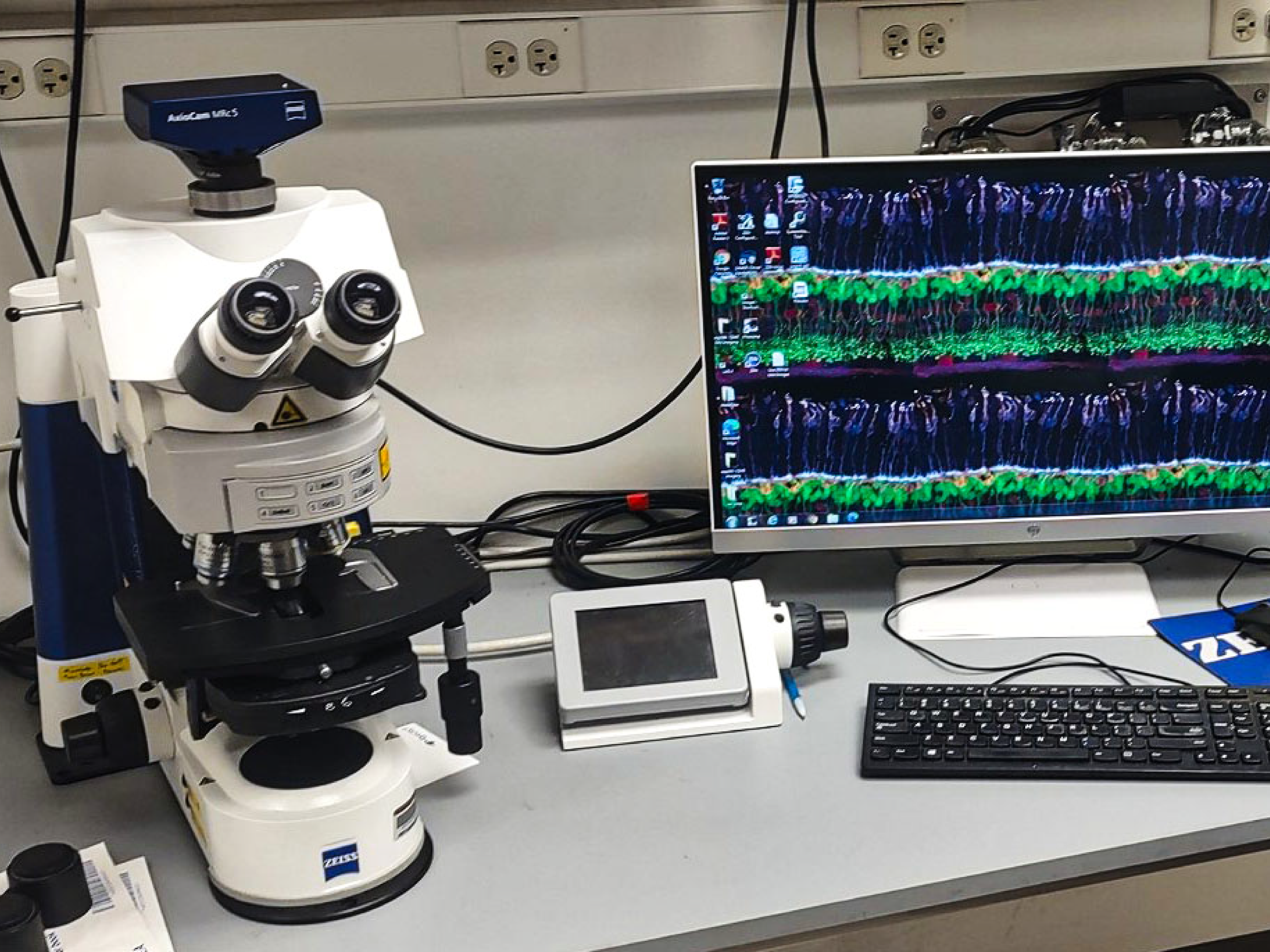
Zeiss Axio Imager Widefield Microscope
 Two
Two 
Expanded equipment access and data gathering on structural and functional analysis
Core A focuses on physiological and structural data gathered from the cellular level (in vitro) to animal models (in vivo) of eye diseases. The work often requires expensive equipment and technical expertise that individual researchers can’t afford but institutions can, such as:
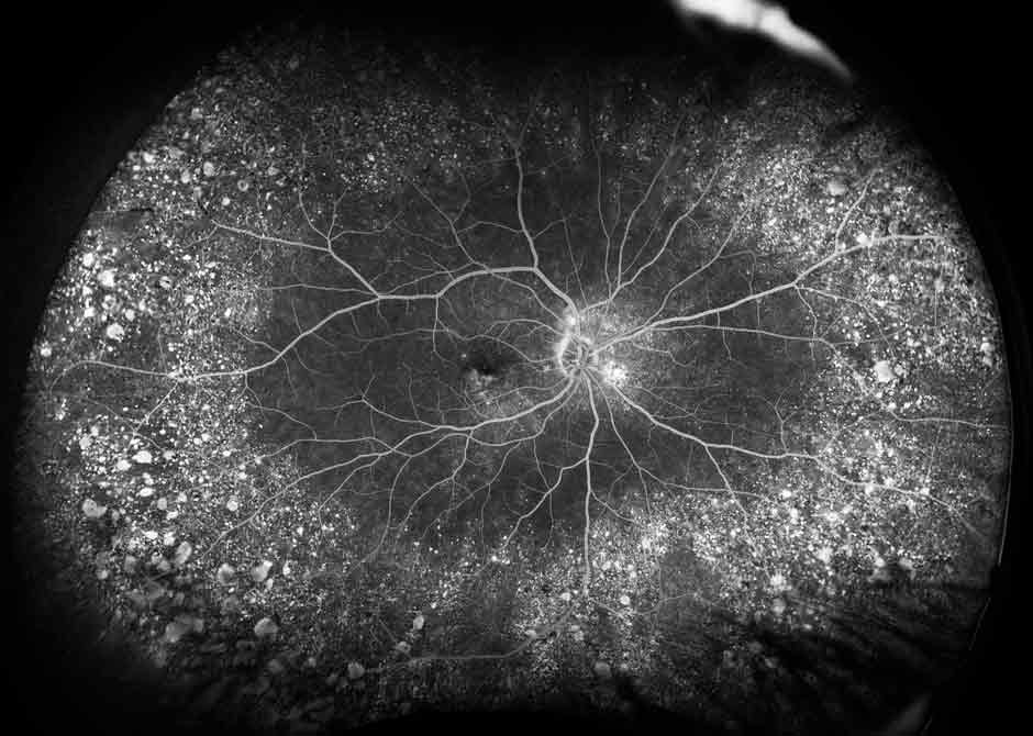
- Angiography
- Electrophysiology
- Fundus photography
- Photocoagulation laser systems
- State-of-the-art microscopes
- Optical coherence tomography
- Tonometry
The Core A team, directed by DOVS faculty Colleen Cebulla, MD, PhD, and Department of Neuroscience faculty Andy Fischer, PhD, will also provide consulting expertise to scientists and labs that need additional analysis or help with developing specific research methods. Such technical support can also assist funded scientists in completing additional studies within their funded projects
For example, when studying macular degeneration, glaucoma or inherited retinal dystrophy, a researcher may study one aspect of a condition, such as histology. The P30 cores offer the ability for the same scientist to expand their work by taking in vivo pictures over time and some electrophysiologic functions of the diseased tissues or eyes.
“This P30 grant will enhance research of funded investigators and also help faculty without grants who need critical pilot data in order to write a strong new grant proposal." - Dr. Moroi.