
Ohio State forms the Institute on Aging to unite breadth of research across disciplines

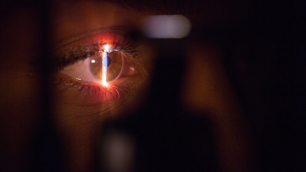 Keratoconus is a progressively degenerative disease in which the cornea, or the transparent, dome-like tissue on the front of the eye, thins and protrudes into a cone shape. These alterations can cause blurry, distorted vision.
Patients with this disease are currently diagnosed using structural imaging techniques such as corneal topography and tomography to detect abnormalities in the eye. But even with this technology, the progression of keratoconus remains difficult for physicians to predict.
Scientists at The Ohio State University College of Medicine and the Ohio State College of Engineering are developing alternatives to current technology that could transform how keratoconus is identified and treated.
Recently, a research team led by Jun Liu, PhD, professor of Biomedical Engineering at Ohio State, confirmed that a high-frequency ultrasound method called ocular pulse elastography (OPE) could help diagnose keratoconus. OPE differs from current techniques because it uses acoustic energy to measure biomechanical properties of the cornea.
“The current techniques — topography and tomography — cannot measure the biomechanics of the cornea. They can’t tell whether the cornea is mechanically weak overall or in some regions,” Dr. Liu says. “They do a great job measuring the shape and thickness, which are useful for diagnosis, but a biomechanical evaluation promises to better diagnose keratoconus.”
Dr. Liu’s newest collaborative work with Andrew Hendershot, MD, clinical associate professor of Ophthalmology and Visual Sciences at the College of Medicine; Xueliang “Jeff” Pan, PhD, clinical assistant professor in the Department of Biomedical Informatics at the College of Medicine; Sunny Kwok, a PhD student in the Department of Biomedical Engineering; and William Liu, a Computer Science and Engineering undergraduate student, revealed specific biomechanical markers measured by OPE that could indicate whether a patient has keratoconus.
One biomechanical parameter they evaluated was corneal axial displacement (CAD), a measure of how much the cornea has moved axially in response to the ocular pulse. A study found that patients with keratoconus had significantly higher CAD compared to normal eyes, and CAD increased as keratoconus progressed over time.
Another metric the research group analyzed was corneal stiffness index (CSI). Patients with keratoconus had a lower CSI than normal patients, and stiffness decreased with keratoconus progression.
The trends in CAD and CSI support the hypothesis that as keratoconus progresses, the cornea experiences mechanical weakening over time. Because they show a correlation with the disease progression, the data also suggests that CAD and CSI could become helpful diagnostic tools for characterizing keratoconus.
“What excites me most is that a quick ultrasound scan may give us information to differentiate normal from keratoconus corneas, and it gives us a biomechanical index that correlates with the disease severity,” Dr. Liu says.
When combined with other methods, the OPE technique could allow physicians to diagnose and manage corneal diseases like keratoconus more effectively.
“Ultrasound is safe and cost-effective, so this method can potentially be routinely used in ophthalmic clinics,” Dr. Liu says. “The biomechanical measurements will add to the clinical workup so the physicians can be better informed to decide whether and when to treat a patient to prevent vision-threatening progression.”
Learn more about this study in the journal PLOS ONE.
This research was supported by the National Institutes of Health.