
Researchers unlock the architecture of the astrocyte network, revealing insight into the astrocyte’s mysterious role in the brain
 Ohio State researchers are delving into the perplexing morphology of astrocytes, a specific type of glial cell located in the brain. Astrocytes are star-shaped cells uniformly distributed across the central nervous system. They are integral to brain function and influence how the brain communicates with the body.
Despite knowing what they are and what they do, there is very little information available about the cellular structure of the astrocyte syncytium.
Min Zhou, MD, PhD, associate professor of Neuroscience at The Ohio State University College of Medicine, led his interdisciplinary research team in their exploration of the cellular morphology of astrocytes.
“The function of astrocytes in the adult brain is still mysterious,” Zhou says. “Seeking answers to our curious neurobiological questions prompted our research team to embark on this seven-year research project.”
Zhou says the team first set out to determine the structural details of the syncytium and how this influences neuronal synapses, the information processing unit of the brain.
“Disruption of the astrocyte network occurs in every neurological disorder including strokes, traumatic brain injuries, epilepsy and depression,” Zhou says. “Our goal is to establish the anatomical structure of the astrocyte network to evaluate potential causal links between altered astrocyte anatomy and disease processes.”
To do this, they utilized a recently available imaging tool called serial block-face scanning electron microscopy, which made it possible to address several long-standing questions about the structural details of astrocytes and the architecture of astrocytic networks, without the cells incurring any damage. This imaging tool was used to obtain cross-sectional images of these three astrocytes, their intracellular organelles, blood vessels and their associated neuronal structures.
Using this form of electron microscopy, the research team generated 500 cross-sectional images of a hippocampal region which, when stacked together, were used to trace the astrocytes, neurites, and blood vessels.
“This required knowledge to identify each structure, as well as the patience and well-trained skillset to trace through each image,” Zhou says. “In addition, the manual tracing to capture the smallest details of the astrocyte structures required exceptional patience as it took a collective effort over many years to complete.”
This study integrated medical research, computational modeling, and art design to illustrate the ultrastructural architecture of an astrocyte network. Recreating the ultrastructural details of the astrocyte syncytium ensures that other researchers can continue to investigate the role of astrocytes in pathologic disease processes.
“Form follows function – now the availability of the ultrastructural architecture of an astrocyte network should facilitate the physiological study of astrocyte function in the normal brain and the pathological involvement of the astrocyte network in various diseases,” Zhou says.
This was was supported by awards from the National Institutes of Health and the National Health Foundation.
Read more about Zhou and his team's research findings in Progress of Neurobiology.
More from Ohio State
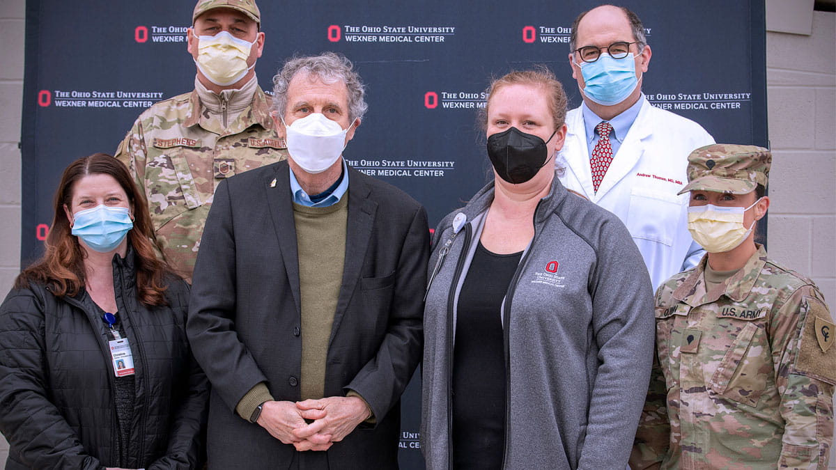
Sen. Brown tours COVID-19 testing site at CAS
Sen. Brown talks with members of the Ohio National Guard who’ve joined health care workers across the state on the front lines in the battle against COVID-19.
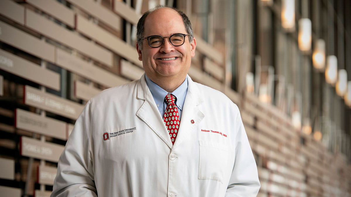
Decades of leadership experience prove vital in the state’s fight against COVID-19
Dr. Andrew Thomas and his decades of leadership experience at The Ohio State University Wexner Medical Center have been vital in the state’s fight against COVID-19.
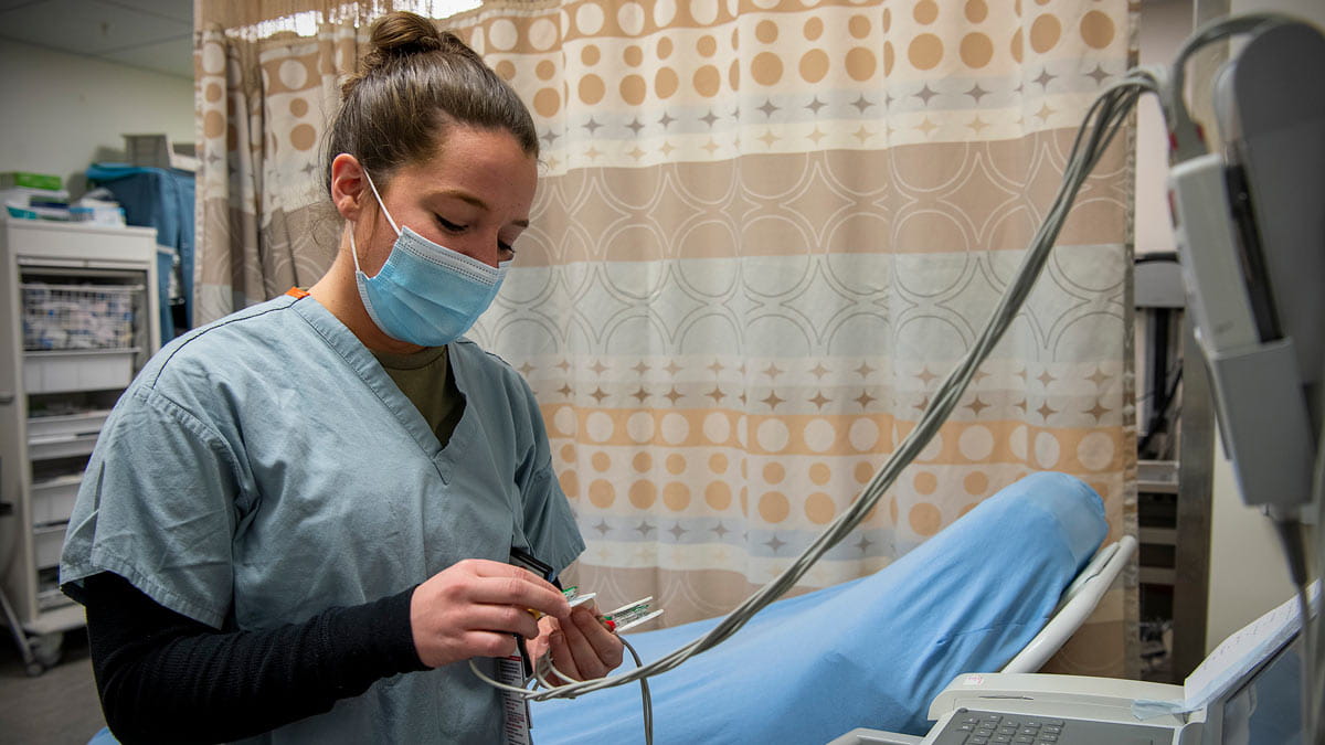
Ohio National Guard joins frontline staff at The Ohio State University Wexner Medical Center
Dozens of other Ohio National Guardsmen have been sent to assist in non-clinical roles, including environmental services, nutrition services and patient transportation. And more are expected in the coming weeks.