10 College of Medicine residency programs recognized among top 20 programs nationwide

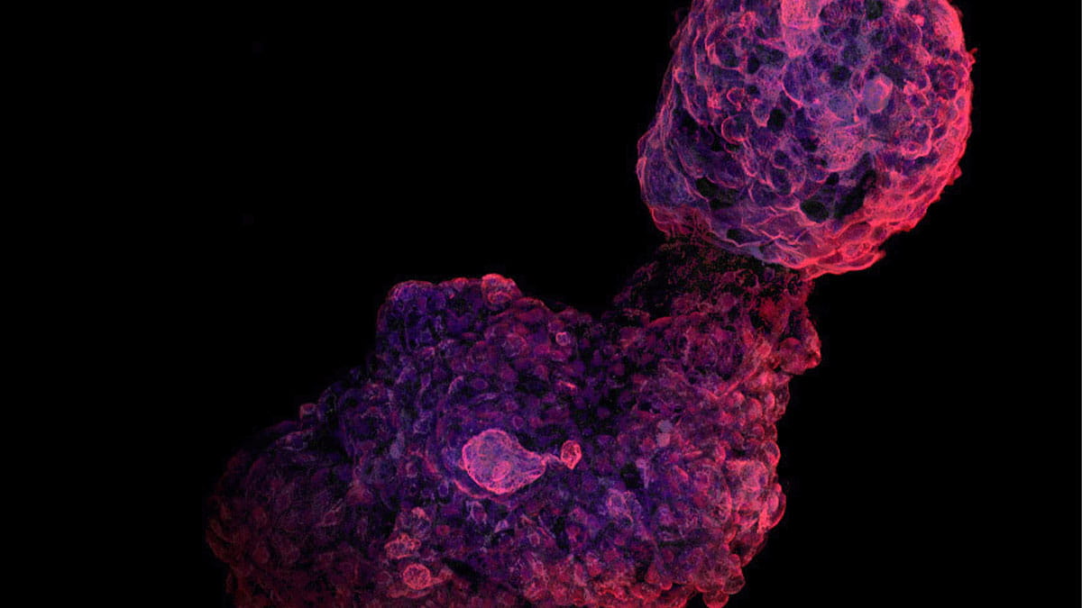
Nikon Small World is regarded as the leading forum for showcasing the beauty and complexity of life as seen through the light microscope. Their 2025 call for submissions to those working across a wide array of scientific disciplines was delivered far and wide. It led three members of a research laboratory at The Ohio State University College of Medicine to submit their piece, called “Human iPSC-derived cardiac organoid.”
Divya Sridharan, PhD, a research scientist in the laboratory of Mahmood Khan, PhD, M.Pharm, professor in the Department of Emergency Medicine at the College of Medicine, and two summer interns, Syed Ashraf and Salvia Zafar, used confocal, deconvolution and image stacking techniques to magnify the beauty and complexity of a human induced pluripotent stem cell (iPSC)-derived cardiac organ grown from stem cells.
It recently received the Image of Distinction Award at the 2025 Nikon Small World Photomicrography Competition. To better understand how they arrived with the elements to capture an image using the Nikon AXR microscope, Dr. Sridharan provided background information on how the work was carried out, and what factors impacted the research and construction of the award-winning image.
Dr. Sridharan said that, for her approach to developing an understanding of the mechanisms underlying the pathological remodeling that takes place after a heart attack, they had to delineate the interactions among different cardiac cell types in a healthy heart versus a diseased heart.
She says that conventionally, human induced pluripotent stem cell-derived cardiomyocytes cultured in tissue culture plates have been used to model the disease. However, the cells do not mimic the changes observed in an actual heart. mainly because the 2D environment alters the behavior of the cells. Then, she figured out how to overcome this limitation.
“I optimized a protocol to culture and differentiate the human iPSCs to functional cardiomyocytes in a 3D microenvironment,” Dr. Sridharan says. “Syed and Salvia participated in culturing and differentiating the cells to 3D cardiac organoids and staining the organoids with cardiomyocyte-specific markers to understand the structural and cellular organization of the cells.”
Dr. Sridharan’s current research focuses on differentiating human iPSCs to cardiovascular progenitors for myocardial regeneration. Ashraf and Zafar. her summer interns. both graduated from Ohio State, and Zafa is now a medical student at the Marshall University Joan C. Edwards School of Medicine, in West Virginia.
Congratulations to this team for this award, and for using their collaborative creativity to create this striking image.