Two College of Medicine students fill spots on University Board of Trustees

The speed of artificial intelligence (AI) and its extensive capabilities across healthcare are breathtaking to us all.
For those of us who work in pathology, AI offers exciting opportunities to help transform primary and clinical diagnosis, enhance treatment decision making and improve patient care.
The Ohio State University College of Medicine’s Department of Pathology was already in the lead in implementing technology to advance that effort. In 2018, the department was the first in the United States to use digital images for primary diagnosis, paving the way for using AI algorithms for diagnostic use.
That undertaking first required us to convert glass slides into millions of pixels — what you might think of as “alphabets” of an image. Once the slide is converted into pixels, it’s AI that can now decipher that image, translating its “alphabet” features to feed into an algorithm that can indicate whether the image is cancer.
The AI algorithm can then potentially determine the grade — and thus, the stage — of the cancer, all within seconds, and can predict, for example, whether a patient will benefit from the currently recommended therapy.
AI’s powerful predictive capability also integrates and assimilates other information in the patient’s medical record to give us that determination.
Digitized images can also be seamlessly integrated into the electronic medical record and the lab information system. All these developments mean pathologists are no longer tied to the microscope. Images can be transferred to the right pathologist for the right diagnosis at the right time.
Currently, we’re at the stage of building our own AI algorithms, testing their use in clinical trials as well as implementing commercially available solutions. Most recently, our team completed clinical trials using AI algorithms to detect prostate cancer, which was commercially available and approved by the FDA. We’re now conducting clinical trials related to breast cancer, where we’ve found that AI can identify cells that mimic benign cancer, and that it can distinguish among the different breast cancer subtypes, for example, ductal, lobular or small cell.
The clinical trials have also shown that AI can not only quantify the amount of cancer in the biopsy and look for cancer features, it can also help measure the distance of margin to the cancer cells to the millimeter — something we used to do by physically placing rulers on the glass slide. An accurate measure of the tumor and distance to margin can make a big difference in identifying the patient’s cancer stage.
These AI tools allow us to make pathology more objective and standardized so that a patient in Altoona, Pennsylvania, or a patient in Chillicothe, Ohio, with the same type of cancer will have more access to more accurate measurements and more objective tools for a personalized diagnosis. We’re now working on developing and implementing AI algorithms for gastric, pancreatic, lung and colon cancers.
Meanwhile, we continue to digitize slides. In December 2025, we had scanned 5 million images, making our department’s collection one of the largest archives of pathology images with patient material, annotations and data in the United States. We also now have a computational pathology group, where PhD scientists are working with pathologists, medical students and graduate students to build more algorithms and predictions.
To boost these efforts, we recently built a model similar to ChatGPT, customized to each case, where a pathologist can converse with the model, asking the computer questions about the patient’s cancer and, for example, treatments this patient might respond to.
Data collection from these efforts will enable us to do more collaborative research across different disciplines at Ohio State, work with startup companies that have unique algorithms to apply them on our data set and collaborate with other academic institutions.
We’re publishing about our work in journals, and we’ve partnered with AI companies that are validating their algorithms at Ohio State.
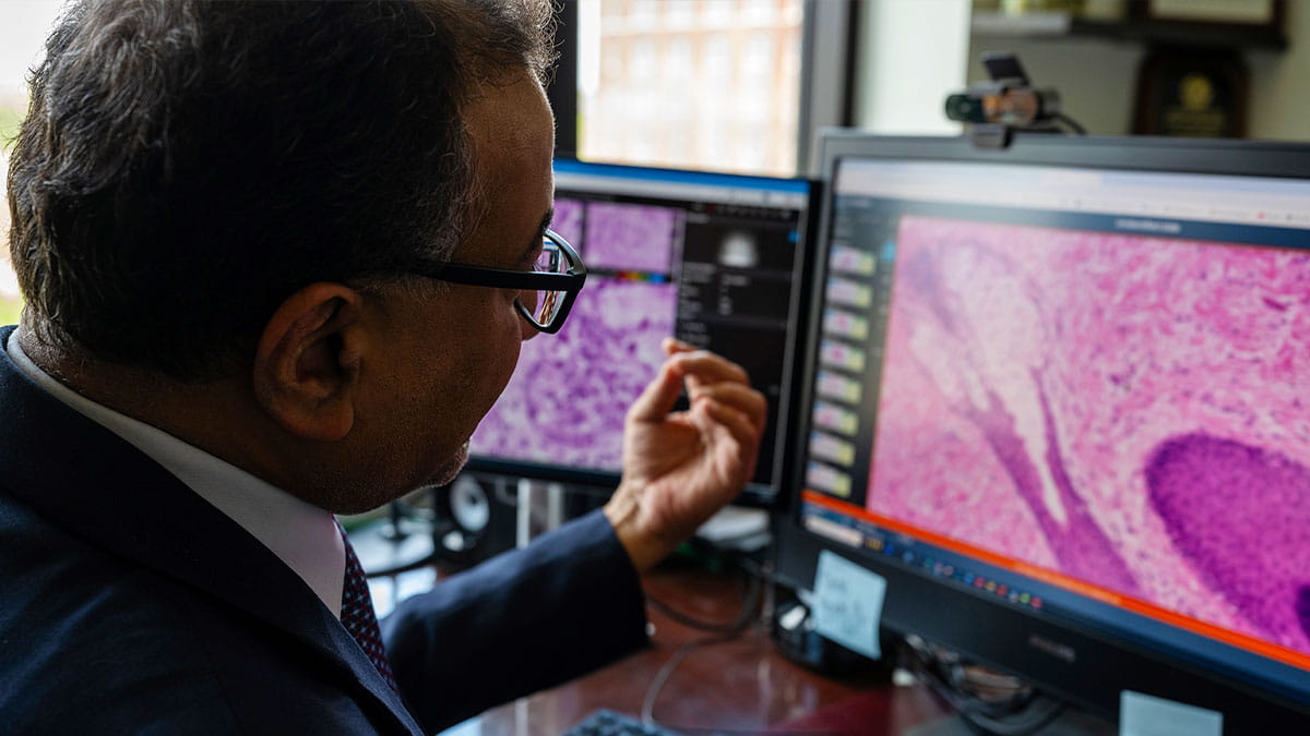
Graduate-level education and training programs are already preparing students, pathology trainees and others in AI’s value for patient care, Parwani says.
We’re also focused on training future doctors, specialists, pathologists and others with this technology.
While all medical students rotate through pathology over their four years, we now also offer electives in AI, in which they work with a faculty member and do research. We’re discussing ideas for incorporating AI into the pathology curriculum. We have already launched an online master’s program in computational pathology, to include AI, and which will be open to medical students beginning fall 2027 or spring 2028.
We have also started a precision pathology fellowship, which includes AI, and is the first in the nation to offer advanced training in diagnostic pathology combined with digital/computational pathology and molecular pathology. Finally, with a grant from AstraZeneca, we’re developing an online program to provide education to students, pathologists and chief hospital medical officers on digital pathology and AI. Many pharmaceutical companies are eager to have pathologists and physicians knowledgeable about AI.
The shortage of pathologists worldwide makes the advances with AI even more impactful. Some countries don’t even have a single pathologist.
AI is also a technology that anyone engaged in health and medical care should be trained in. Whether you’re a medical student in Africa or a pathologist in Columbus who’s also covering a hospital in Wooster, Ohio, your AI knowledge will be invaluable.
With this technology, we can help so many more patients. Truly, AI in medicine and pathology is just the beginning of a very exciting and transformative journey.