Michael Prats, MD, selected for Columbus Business First's 2026 40 under 40

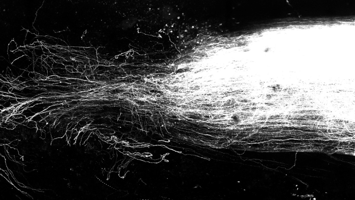 Capitalizing on the flexibility of tiny cells inside the body’s smallest blood vessels may be a powerful spinal cord repair strategy, new research suggests.
In mouse experiments, scientists introduced a specific type of recombinant protein to the site of a spinal cord injury where these cells, called pericytes, had flooded the lesion zone. Once exposed to this protein, results showed, pericytes change shape and inhibit the production of some molecules while secreting others, creating “cellular bridges” that support regeneration of axons – the long, slender extensions of nerve cell bodies that transmit messages.
Researchers observed axon regrowth in injured mice that received a single treatment injection of the growth-factor protein, and the animals also regained movement in their hind limbs. An experiment involving human cells suggests the results are not restricted to mice.
“There’s a lot more that can be learned and a lot that can be expanded, but the more we worked on this, the more stunned we really were by the potency of this single treatment and how effective it was,” said senior study author Andrea Tedeschi, PhD, associate professor of neuroscience in The Ohio State University College of Medicine. “This finding goes beyond spinal cord injury – it has implications in brain injury and stroke, and neurodegenerative diseases as well.”