Research results in further understanding of the mechanisms of protein-mediated DNA annealing
Twenty years ago, researchers discovered that the protein λ-Redβ exhibited unusual DNA binding properties. The most studied protein, λ-Redβ binds weakly to single-stranded DNA, not at all to pre-formed double-stranded DNA and tightly to a duplex intermediate of annealing formed when two complementary oligonucleotides are added to the protein sequentially.
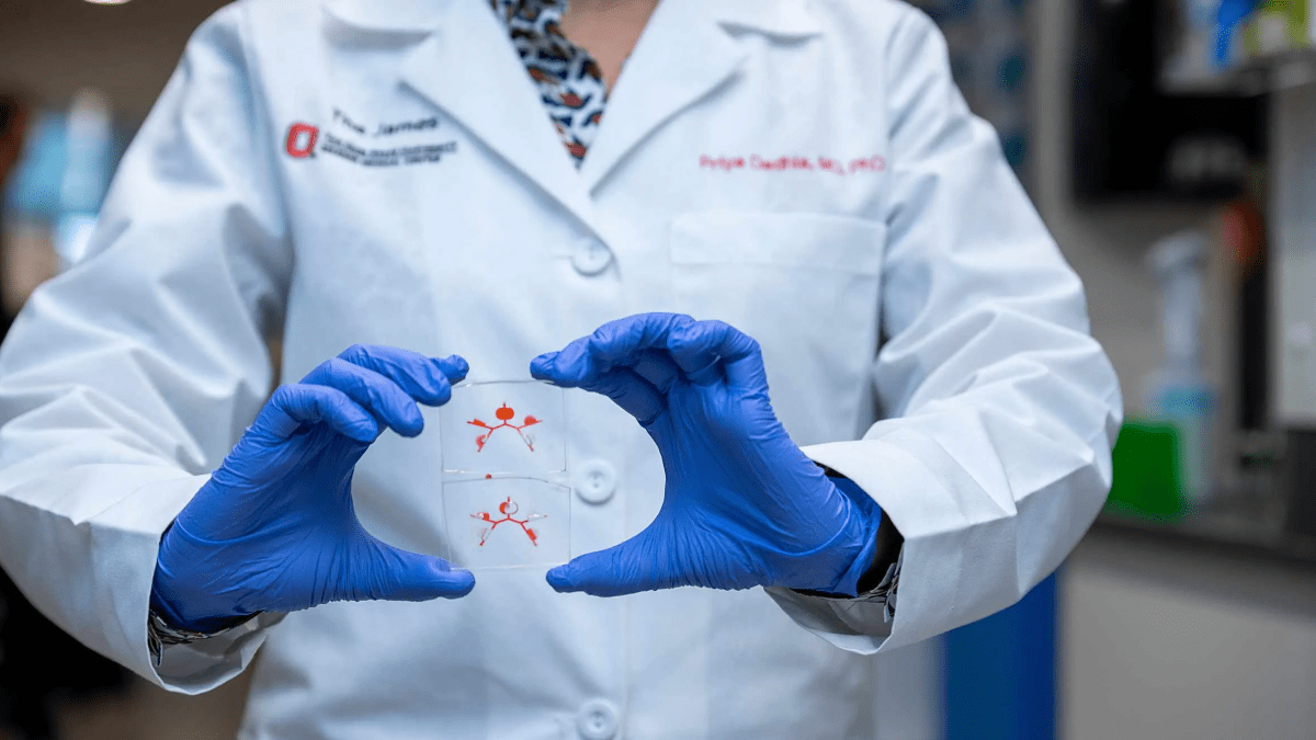
Charles Bell, PhD, associate professor of Biological Chemistry and Pharmacology at The Ohio State University College of Medicine, has dedicated his career to understanding how proteins like λ-Redβ affect annealing, the process of joining of single-stranded DNA or RNA by hydrogen bonds to form a double-stranded polynucleotide.
A new study, “Structure of a RecT/Redβ family recombinase in complex with a duplex intermediate of DNA annealing” just published in Nature Communications.
In the study, Dr. Bell and his team of researchers used single particle cryo-electron
microscopy (cryo-EM), which is a method for imaging specimens in their native state, without the use of dyes or fixatives. The method facilitates the study of fine cellular structures, viruses and protein complexes on a molecular level.
This allowed the researchers to “see” a structure of λ-Redβ, which revealed a helical protein filament bound to a DNA duplex that is highly extended and unwound.
“We discovered λ-Redβ shares a common conformation with RAD52 along with a similar mode of single-stranded DNA-binding,” Dr. Bell says. “These data provide insights into the mechanism of protein-catalyzed single-stranded DNA annealing to repair double-stranded DNA breaks.”
Dr. Bell furthers that this research builds on his inquiry into the mechanisms at play in allowing proteins to form large structures without increasing genome size. By studying protein function in this way, soon they will be able to translate their findings into studying function in cells.