NIH funds support emerging study of machine learning to aid in measurement of blood oxygenation status
Measurement of blood oxygen saturation, or the percentage of hemoglobin that carries oxygen in blood, has long been employed as a diagnostic tool to guide therapy and intervention. This is because it provides valuable information about blood circulation and the body’s oxygen supply and demand status.
While the commonly used finger pulse oximeter only measures arterial blood oxygen saturation, doctors need to know the oxygen levels at different locations within the heart in order to treat certain heart conditions. Currently, these measurements are made in a cardiac catheterization laboratory under fluoroscopic guidance. Although considered the gold standard, this method has significant drawbacks. The procedure is invasive; the catheter has the possibility of damaging already vulnerable tissue, and the patient may have to undergo sedation to ensure proper placement. There is also the risk of over-exposure to radiation as X-rays are used for catheter guidance.
In an attempt to mitigate the risks associated with the traditional fluoroscopy approach, the use of noninvasive magnetic resonance imaging (MRI) has been explored. This eliminates radiation exposure and the need for sedation. MRI signals in blood are sensitive to oxygen saturation, but historically, the multiparametric Luz-Meiboom regression model used to determine oxygen saturation from MRI data has yielded results not suited for clinical application. The margins of error are large as the model does not capture all the relative phenomena and can also have multiple mathematical interpretations.
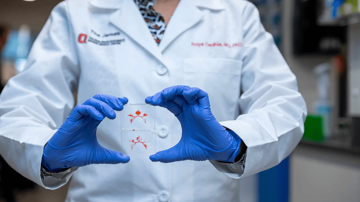
In an effort to create an accurate and precise technique for measuring blood oxygen saturation in the heart, with minimal exposure to radiation, Juliet Varghese, PhD, research scientist at The Ohio State University Dorothy M. Davis Heart and Lung Research Institute, proposes the use of machine learning to analyze the results of MRI scans. The machine-learning algorithm will be pre-trained using MRI data simulated from the Luz-Meiboom model and then augmented with in-vivo data through transfer learning. It is hypothesized that the machine learning-based approach will provide greater flexibility to map the true relationship between magnetic resonance blood signals and oxygen saturation.
During the development of the machine-learning algorithm, Dr. Varghese will simultaneously develop a 3D MRI data acquisition method to generate a volumetric map of oxygen saturation throughout the vascular system. Once both the machine learning and MRI mapping approach are thoroughly developed and validated, they will be used together during a small-scale clinical trial on patients who need to have their heart blood oxygen saturation measured.
“Ideally, we would have a mixture of patients with normal and complex blood circulation,” says Dr. Varghese. “Normal circulation patients, such as those with heart failure, and complex circulation patients having heart defects will allow us to better validate our technique.” These patients will undergo an MRI of the heart and the images will then be passed through the machine-learning algorithm to determine information about blood oxygenation status.
The grant from the National Institutes of Health to fund this work is categorized as a Trailblazer R21 Award. These particular awards offer opportunities for new and early state investigators to pursue research of high interest to the National Institute of Biomedical Imaging and Bioengineering, which funds research at the interface of the life sciences with engineering and the physical sciences.
The funding of a machine-learning algorithm for MRI-based blood oxygen saturation will allow Dr. Varghese and her team to explore noninvasive techniques for attaining biometric data. Successful implementation of a noninvasive accurate technique for mapping cardiac blood oxygenation would provide a diagnostic method that is safe and accurate, and can be repeated without concern of side effects or radiation exposure.
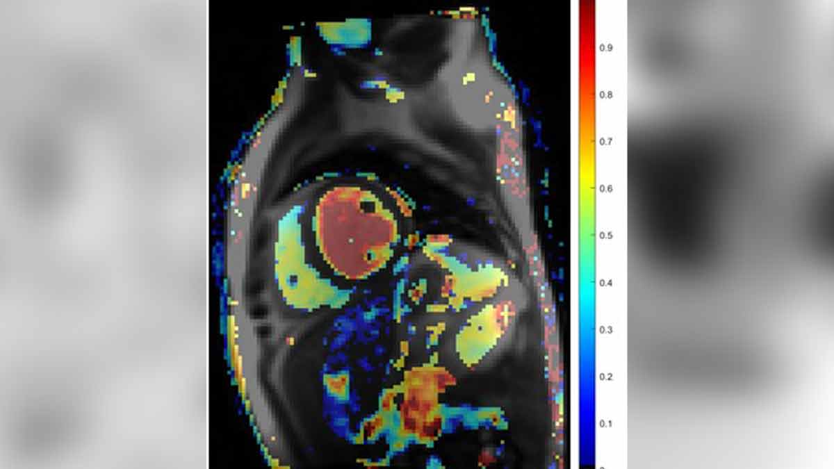
This is a blood oxygen saturation map overlaid on an MRI image of the heart. In a healthy individual, arterial saturation ranges between 95 — 99%, while venous saturation ranges from 60% – 80%. In this example from a heart failure patient, the right ventricle shows low venous oxygen saturation while the left ventricle shows normal arterial saturation levels, indicating impaired oxygen delivery.