Alumni Society presents awards recognizing distinguished career achievements in practice and teaching of medicine

AI’s capabilities advance the detection, diagnosis and treatment of cancer.
Imagine that you’re a patient waiting for imaging results — for answers and a clear next step. If you’re facing the possibility of cancer, what you want most is expert reassurance.
For people undergoing cancer screening today, that reassurance may involve knowing that their care team is pairing their expertise with tools like artificial intelligence (AI) to review scans and find subtle patterns the human eye can’t reliably spot — tools that will help them detect disease earlier and tailor treatment with greater confidence.
At The Ohio State University College of Medicine, researchers are developing algorithms for AI to give them better tools to improve their data-driven decision making in cancer diagnosis, care and treatment.
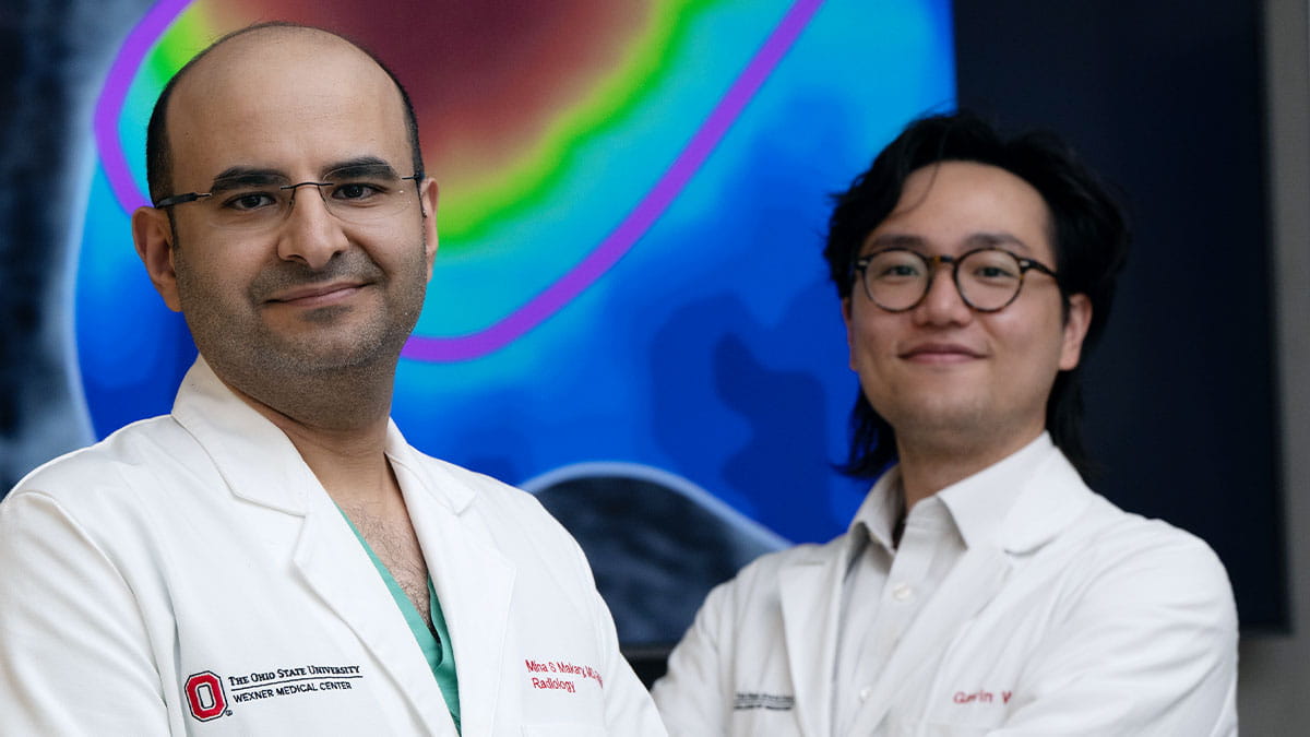
Mina Makary ’13 MD, ’17/19 Res, associate clinical professor of Radiology in the Division of Vascular and Interventional Radiology, is a national leader in this arena. A vascular and interventional radiologist, Makary has been developing algorithms — sets of instructions a computer learns from to obtain and mine imaging data — to provide better disease evaluation and prediction models. He trains AI by feeding it curated datasets, validated patient evaluations and scans, and adjusts internal parameters until it performs more accurately than manual predictions.
The result: AI is able to accurately identify what’s wrong in an image, computed tomography (CT) scan or magnetic resonance imaging (MRI), allowing for improved diagnosis and planning for next steps.
“AI offers powerful analysis, which detects different features that go beyond the abilities of the human eye,” Makary says. “If we can see findings better, we can characterize them better and then we can diagnose and predict treatment response better.”
Radiogenomics is an emerging discipline that combines the fields of radiology and genomics, examining the relationship between a disease’s imaging features and its molecular characteristics. It focuses on how imaging data can correlate with a patient’s medical information by integrating quantitative data from medical images with a patient’s physical manifestation of disease and clinical outcomes.
AI analyzes a CT or MRI, identifies unique features and can even predict tumor biology size and determine the most effective treatment or chemotherapy regiment. This could change uniform treatment into personalized care and improve response rates, Makary says.
“These AI applications are possible without conventional invasive tissue sampling, which potentially spares patients from biopsy procedures and their associated risks,” he says.
Building on his research interests in AI applications in imaging, Makary worked on the first-ever study in the country that curated abdominal CT scans and presented an algorithm and computational framework for automated detection and characterization of inferior vena cava filters, including in patients with cancer. The aim was to identify implanted filter devices in patients and support the safe removal of those filters to prevent long-term complications and improve outcomes. Still in the research phase, this advancement confirms that AI supports clinicians but will never replace them, he emphasizes.
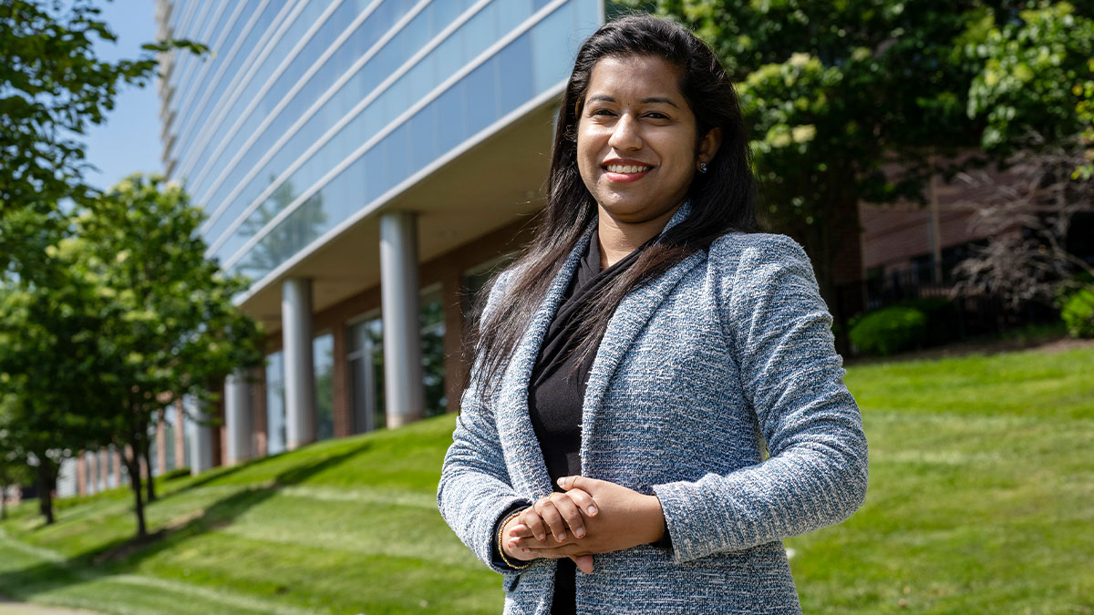
Oncologist Arya Roy, MD, is working with AI experts to build a risk prediction tool that will predict recurrence of lobular breast cancer after treatment.
Simeng Zhu, MD, assistant clinical professor of Radiation Oncology, studies how AI can help care teams treat the cancers he sees most often, including brain, spine and head and neck cancers. His team uses AI to analyze medical images and help predict how a person may respond to treatment. Zhu says the key is AI’s ability to quickly review substantial amounts of information, including scans and lab reports gathered during diagnosis.
“We don’t make decisions based on one data point, but on multimodal data, combining scans, lab results and clinical notes,” Zhu says. “AI can help relieve some of the burden on our healthcare providers, streamline the whole process and allow us to integrate knowledge into new discoveries and treatments.”
For example, he says, AI has the potential to quickly pick out areas of interest (such as a lung nodule) for diagnostic radiologists to further analyze, and AI has already been deployed in many radiation oncology departments across the world — including Ohio State’s — to help accelerate the radiation treatment planning process without compromising quality.
“Now we develop AI algorithms that can generate a rough assortment of segmentations — the contours and shape of the tumor and the normal tissues,” Zhu says. “All we need to do is modify them instead of drawing everything from scratch.
AI’s ability to accurately analyze images improves diagnosis and treatment planning, says Mina Makary, ’13 MD, ’17/’19 Res, an interventional radiologist.
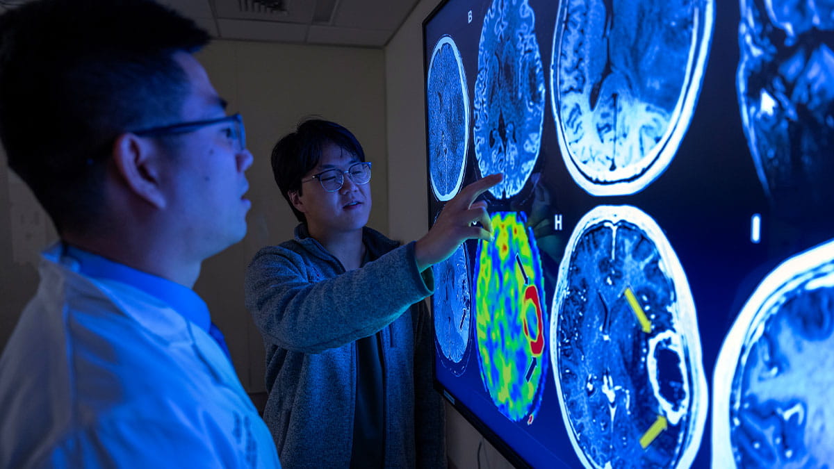
Simeng Zhu, MD (left), is focused on research exploring AI’s ability to identify tumor and tissue shape. First-year medical student Michael Kong is helping with the research.
After receiving undergraduate degrees in biomedical engineering and computer science, Michael Kong, M1, now works with Zhu on auto-segmentation, the process of automatically identifying and delineating different objects within an image. They can “feed” a brain MRI to an algorithm they trained to automatically draw the locations of metastases in the brain, leveraging AI’s strengths with pattern recognition.
“That’s the disease that we’re trying to have the computer label. That way it catches all the brain metastases no matter how many there are,” Kong says. “In practice, this could mean fewer repeat scans and quicker decisions about next steps.”
This research keeps them at the forefront of understanding how to use AI in clinical spaces. Other researchers are even examining the use of AI to predict whether patients will have a good response to a particular therapy.
“Having an AI algorithm that can stratify the patient’s outcome based on whether it’s going to succeed with a standard treatment could allow an intensified approach,” Zhu says. “Or, it could reduce standard treatments for some patients we could be overtreating.”
After ductal cancer, lobular breast cancer is the second most common type of breast cancer. It can be harder to spot on a mammogram because it often grows in thin lines rather than forming one clear lump, says Arya Roy, MD, assistant clinical professor of Internal Medicine in the Division of Medical Oncology at The Ohio State University Comprehensive Cancer Center – Arthur G. James Cancer Hospital and Richard J. Solove Research Institute.
Roy specializes in caring for people with this hard-to-detect form of breast cancer.
“Lobular breast cancer presents in this puzzling pattern, so it’s often not detected until it spreads or becomes large,” Roy says. “It typically doesn’t respond to chemotherapy and can recur even after 10 years.”
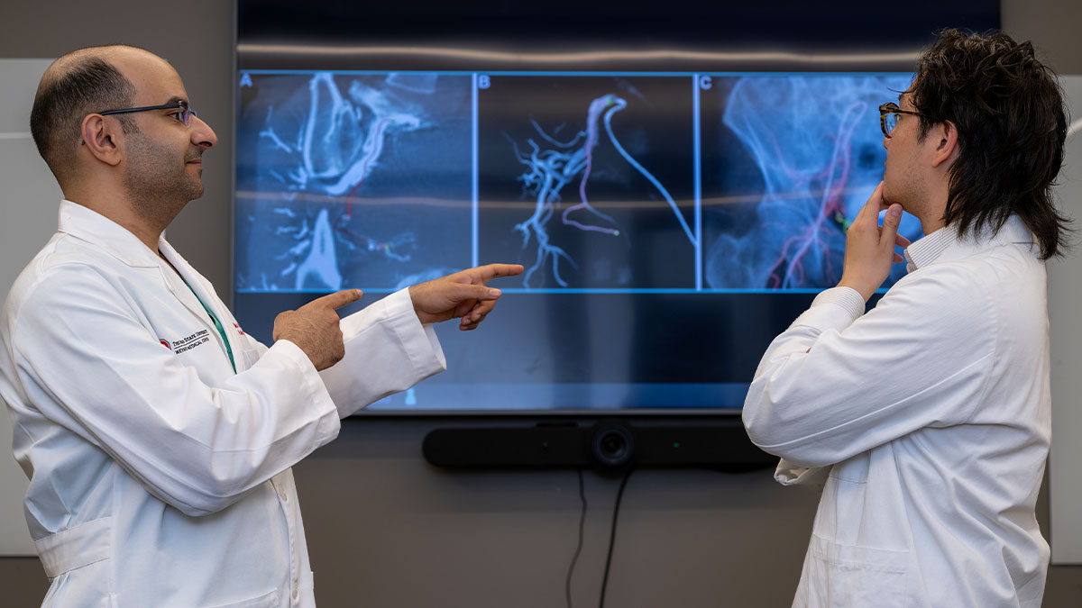
AI’s ability to accurately analyze images improves diagnosis and treatment planning, says Mina Makary, ’13 MD, ’17/’19 Res, an interventional radiologist.
A screening tool to predict recurrence of ductal cancer after initial treatment exists but doesn’t translate effectively for lobular cancer. So, Roy is working with a team of AI experts, led by Khalid Niazi, PhD, MS, associate professor of Pathology, to build an actual risk prediction tool.
Niazi’s research lies at the intersection of AI and pathology. His team is developing tools to identify groups of cells and specific patterns they see in digital pathology slides. As part of this process, traditional glass tissue slides are digitized, then converted into high-resolution images analyzed by special software.
This, with Roy’s clinical input and determining where they need AI’s help, informs their work to develop an AI model that combines clinical features of patients and biomarkers that are present in pathological images. The goal is to pinpoint recurrence and metastatic patterns that will enable clinicians to accurately identify people who are more likely to have recurrence in five years, 10 years and beyond initial treatment.
“After training and teaching the model, it will go through validation with a bigger cohort so we can make sure that the data or the tool that we are developing is accurate,” Roy says.
For now, oncologists must continue to rely on antihormonal treatment options and trust their patients to alert them to any new symptoms that arise to help identify cancer recurrence.
“This is a space that actually needs a lot of research so we can make better and personalized treatment options for these patients,” Roy says.