Featured expert
- Irene Mynatt, DO, associate professor of Emergency Medicine at The Ohio State University College of Medicine.


Throughout her career, Margaret Ginn-Pease, PhD, dived into research on biochemistry and human cancer genetics and loved teaching and mentoring students as they furthered their paths of discovery. She believed anyone could be a scientist if they were open to stretching their mind, asking questions and remaining curious. When she passed at age 57, her husband, William Pease, MD, who is a recently retired Professor Emeritus of Physical Medicine and Rehabilitation at The Ohio State University College of Medicine, started a fund in her honor, to ensure that her passion for getting young students excited about science lived on.
He worked with College of Medicine staff members to develop the Margaret Ginn-Pease Medical Research Day, an annual two-day event that provides middle school students with hands-on activities, interactions with health sciences professionals and time to explore organs and specimens in the college’s state-of-the-art anatomy laboratory.
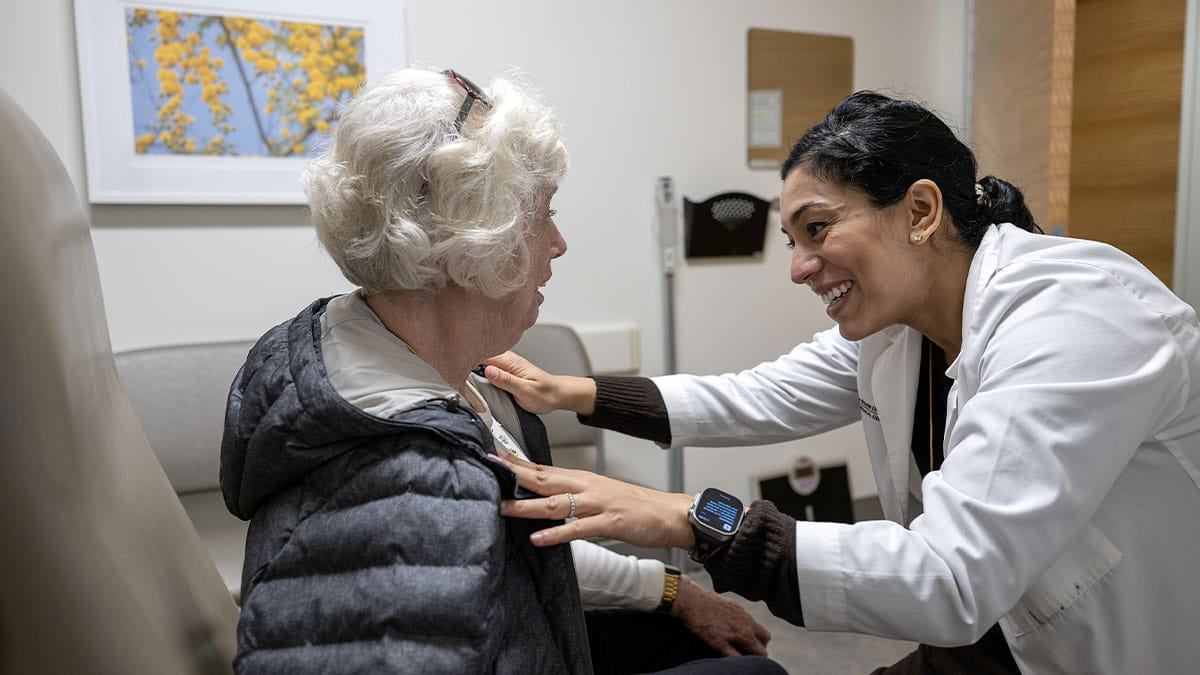
Irene Mynatt, DO, associate professor of Emergency Medicine at the Ohio State University College of Medicine, led this year's event. She prepared the seventh graders from Champion Middle School who attended the event beforehand by visiting them at their school once a month to engage in science experiments. Together, they isolated the DNA of strawberries, created models of pumping hearts and lungs with balloons and empty water bottles and learned how to use stethoscopes and reflex hammers.
Dr. Mynatt says she finds joy in sharing her love of science with the students and that they inspire her to be more creative in coming up with exciting experiments.
At the event, the Champion Middle School students started the program in the college’s anatomy laboratory where they rotated through four stations exploring human organs and specimens, viewing and touching them. Staff, volunteers and health sciences and medicine learners guided them through:
At the end of the second day, there was a special ceremony where Drs. Mynatt and Pease presented the students with white coats, mirroring what medical students receive when they enter the medical profession. Dr. Pease said that his late wife would have been so happy to see the students receive recognition for their exploration of science and get the chance to learn about different career paths that lead to safe, secure professions.