
The Main Campus Imaging Core provides training, expertise and equipment to perform fluorescence confocal microscopy. This facility provides well-maintained point-scanning confocal and multiphoton microscopes and provides training to users to establish independent imaging experiments.
Core Director
Andy Fischer, PhD
Andrew.Fischer@osumc.edu
Core Manager
Lisa Kelly
Lisa.Kelly@osumc.edu
Leica confocal
The Leica SP8 is an upright point-scanning confocal suitable for routine confocal imaging of fixed specimens. This microscope is only available to the laboratories of faculty with a primary or joint appointment in the Department of Neuroscience.
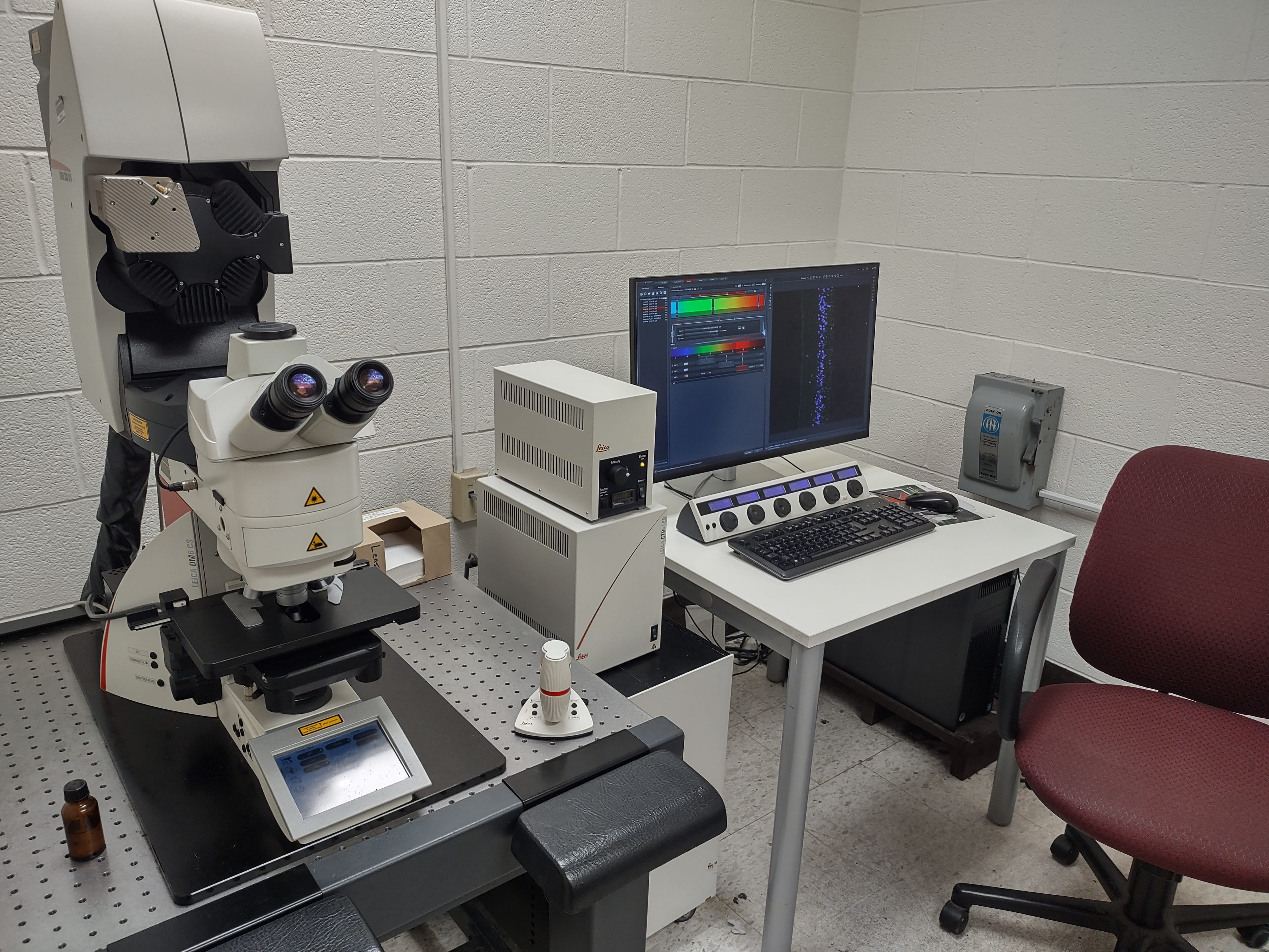
Features:
- Upright fully motorized with 10X, 20X, 40X and 63X high NA objectives
- 40x and 63x are oil immersion lenses
- Motorized XYZ stage with XY stitching
- 4 excitation lines (405, 488, 561 and 640 nm) from solid state lasers and AOTFs
- Adjustable filters for emission spectra
- 2 emission PMTs, 1 HyD emission detector, and 1 transmitted light PMT
- Digital deconvolution
- LASX suite for Z-stack projections, rotations, orthogonal projections and rotation movies
Following in person training on the system users will be granted access to the online calendar to reserve the system and card swipe access to the shared equipment room in Graves Hall. Please note that this is a heavily used shared resource. Thus, during normal working hours reservations are restricted to 4hr blocks of time per lab.
Location: Graves Hall, Rm 4029D
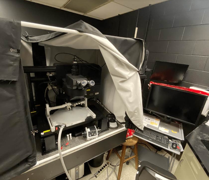
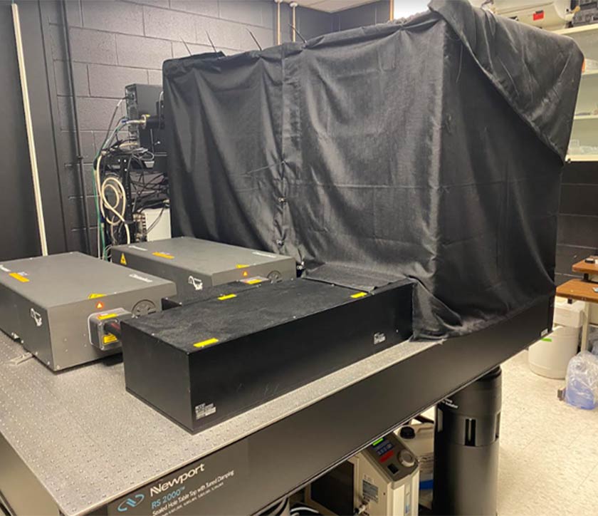
Olympus multi-photon
The Olympus FVMPE-RS is an upright multi-photon microscope capable of confocal fluorescence imaging deep into tissue in brain slices, anesthetized or unanesthetized mice.
Features:
- Dual scan head
- MPE Objectives: 60X dipping, 25X immersion, 20X air and 5X air (MPE Objectives are designed for deep imaging-up to 8 mm working distance/depth)
- Detectors: PMT and GAASP
- High-Speed Scanning at up to 438 Frames-per-Second
- Independent Photostimulation Control
- Microsecond Timing for Electrophysiology and Optogenetics
- Automated Laser Alignment
- FluoView Software
- Coherent Twin Chameleon laser system: 700-1060 nm
- Prior XY motorized stage
- Neurotar Home Cage Arena (for in vivo imaging of unanesthetized mice).
Location: Graves Hall, Rm 4024B
High content cell screening and analysis workstation
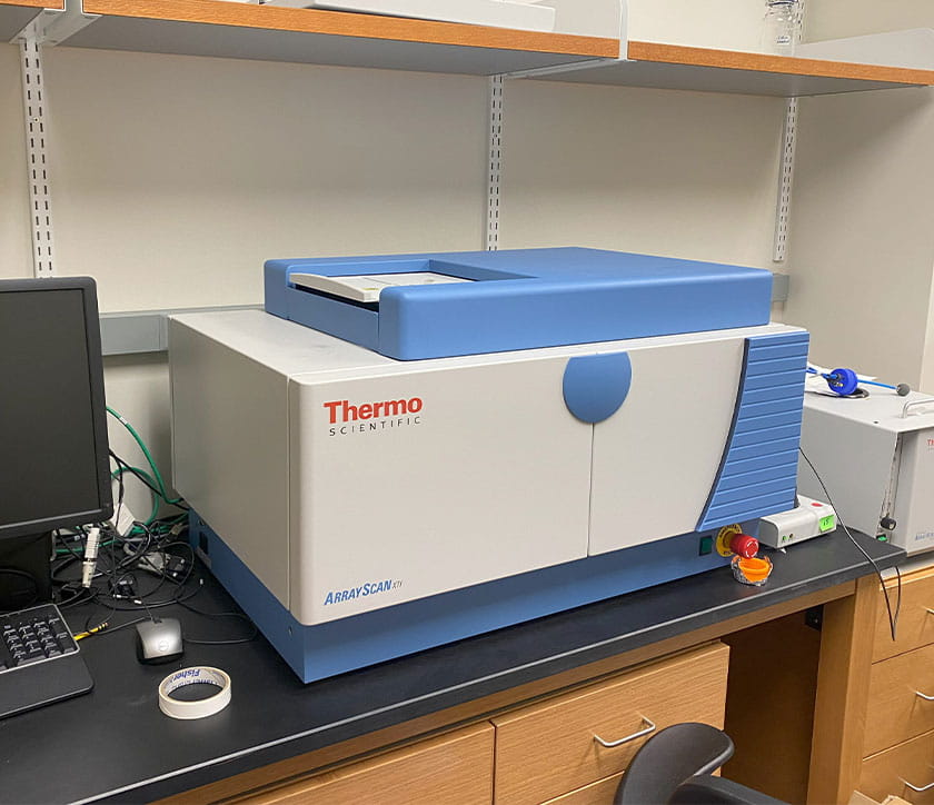 The Thermo Scientific Cellomics ArrayScan HCS high content cell screening and analysis workstation is for high throughput analysis of cell morphology, fluorescent protein expression, live/dead assays, live cell imaging, neurite tracing, etc.
Location: Biomedical Research Tower, Rm 631
User fees
Consultation and training: No charge
Department of Neuroscience users:
- Unassisted use: $25/hr
External users (academic and non-academic):
- Unassisted use: $90/hr
- Assisted use: $180/hr
For multiphoton and high content cell screening and analysis workstation fees, please email the Director.