
The West Campus Imaging Core provides neuroscience researchers with the training, expertise and equipment that they need to perform fluorescence confocal microscopy of cells, tissues and zebrafish embryos in the living and fixed state. We provide access to well-maintained wide-field, point-scanning and spinning disk confocal microscopes, as well as expert consultation and assistance with design and execution of imaging experiments.
Core Director
Anthony Brown, PhD
Anthony.Brown2@osumc.edu
Core Manager
Paula Monsma
Monsma.1@osu.edu
614-293-0939
Location
Rightmire Hall
Available services
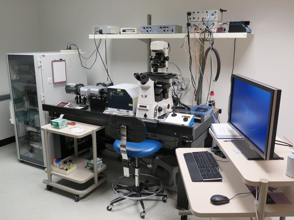 The Andor Revolution WD is an Imaging Core’s spinning disk laser confocal system, ideal for live imaging of cells, tissues and zebrafish embryos.
Features include:
- Inverted Nikon TiE microscope, fully motorized
- Nikon Perfect FocusTM focus stabilization system
- High Numerical Aperture (NA) oil and water immersion objectives
- Differential interference contrast microscopy
- Okolab stage-top incubator with temperature and (CO2) control
- Okolab objective heater
- Motorized XYZ piezo stage
- Two Andor Ultra EMCCD cameras
- One Andor Neo sCMOS camera
- Simultaneous two-color imaging (GFP/RFP or CFP/YFP)
- Yokogawa CSU-W1 fully motorized confocal scanner unit
- Efficient bypass mode allows for wide-field imaging
- Six laser lines: 405nm, 445nm, 488nm, 515nm, 561nm, 640nm
- FRAPPA laser-galvo scanner
- FRAP, FRET, photoactivation and photoconversion applications
- MetaMorphTM Premier software for image acquisition and analysis
Learn more information about objectives and lasers.
Learn more with a detailed description.
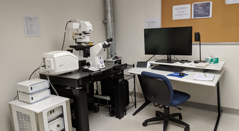 The Zeiss LSM 900 Airyscan 2 is an Imaging Core’s point-scanning laser confocal system, ideal for imaging of fixed specimens.
System features:
- Zeiss Observer 7 fully-motorized inverted microscope stand
- High Numerical Aperture (NA) oil and water immersion objectives
- Four laser lines: 405nm, 488nm, 561nm and 640nm
- Variable secondary dichroic mirrors for detection and cross-talk optimization
- Two high sensitivity GaAsP detectors with variable pinhole sizes
- One Airyscan “superresolution” detector
- Freely rotatable scan area
- Motorized stage
- Ability to scan user-defined regions of interest
- Differential interference contrast optics
- One transmitted light detector
- Zen Blue 3.0 software package
- Multiplex module for faster parallelized image acquisition
- Sample Navigator module for improved sample navigation
Click here for information about objectives and lasers.
Click here for a detailed description.
Technical Support
Your first contact for technical support should always be the Facility Manager, Paula Monsma.
Microscopy Education
You must attend a training seminar to use the microscope. Seminars are held in Rightmire Hall. Please contact the imaging core manager, Paula Monsma, at 614-293-0939 or monsma.1@osu.edu for the seminar schedule.
Andor Revolution WD Manuals and Handouts
Andor Training Handout: Downloading a copy of the handout does not override the need to attend a training seminar.
Andor Microscope Manual: Click here for a copy of the manual located in the confocal room.
Zeiss LSM 900 Airyscan 2 Manuals and Handouts
Zeiss LSM900 Training Handout: Click here for a copy of the handout, for your convenience. However, you will still need to attend a training seminar.
Microscope Scheduling
Scheduling for each instrument is coordinated online. Please contact the Facility Manager, Paula Monsma, at 614-293-0939 or monsma.1@osu.edu for access to the scheduling calendar.
Access Policy
Trainee: Supervised, Monday – Friday, 9 a.m. – 5 p.m.
- Required experience: Seminar and minimum of two hands-on training sessions. Advancement decided on a case-by-case basis.
Standard User: Unsupervised, Monday – Friday, 9 a.m. – 5 p.m.
- Required experience: Seminar and minimum of 10 unsupervised sessions. Advancement decided on a case-by-case basis.
Experienced User: Unsupervised, Monday – Friday, 9 a.m. – 5 p.m., as well as evenings and weekends
- Required experience: Advancement decided on a case-by-case basis.
User fees
Consultation and training: No charge
Ohio State University users:
- $35/hr 9am-5pm
- $30/hr after hours
- $280 flat daily rate for >8hrs
- $35/hr surcharge for assisted use
External users (academic and non-academic):
- Unassisted use: $90/hr
- Assisted use: $180/hr
West Campus core policies
Purpose
To provide neuroscience researchers with well-managed and well-maintained confocal microscopes for imaging of cells, tissue and embryos in the living and fixed state, and to provide expert training, consultation and technical support for the use of this equipment.
Initiation of projects
The core is available to neuroscience researchers on campus. New users should contact the Core Manager to discuss their eligibility for training and access. All researchers on campus are welcome to use the core services but if scheduling conflicts arise, priority will be given to projects in the Department of Neuroscience.
Services
Training, oversight and day-to-day management is provided by the Core Manager, who is supervised by the Core Director. We provide access to the Zeiss LSM 900 Airyscan2 inverted confocal microscope and Andor Revolution WD Spinning Disk confocal microscope, as well as hands-on one-on-one basic training to all new users, and application-specific advanced training to experienced users. First-time users must attend a mandatory training seminar, mandatory one-on-one training sessions, and finally supervised use before being given access to the microscopes. After satisfactory completion of this training, users are given access Monday-Friday 9am-5pm. As users become more experienced, they are given access 24/7, at the discretion of the Core Manager. We also provide consultation and assistance in the development and execution of specific imaging projects, such as those involving live cell imaging in tissue slices, developing zebrafish embryos and cultured cells, and in the processing and analysis of acquired images.
Use of the confocal microscopes can be scheduled in advance using a Google calendar. Entry to the microscope rooms is controlled by keycard. Availability is on a first-come, first-served basis. A lab is permitted to book up to 12 hours each week on each instrument as far in advance as it wishes, but it may not book more than 12 hours in any single week (Sunday through Saturday) until Friday of the preceding week. Users of the Zeiss Airyscan are not permitted to schedule more than 4 hours in one day without permission from the Core Manager, unless they are scheduling on the day of use. Any of these restrictions can be waived by the Core Manager, with appropriate justification. The Core also offers access to a Zeiss Axiophot upright wide-field epi-fluorescence microscope.
User responsibilities
The Neuroscience Imaging Core must be acknowledged in every publication arising from work supported by the Core. For work using the spinning disk confocal, users must also acknowledge NIH S10 OD010383 shared instrumentation grant, which was used to purchase this instrument. For work using the Zeiss Airyscan confocal, users must acknowledge NIH S10 OD026842 Shared Instrumentation Grant, which was used to purchase this instrument. The Core reserves the right to deny access to Users that do not adhere to Core policies or that fail to exercise sufficient care in the use of the microscopes.
Selected publications
Liu Y, Kwok W, Yoon H, Ryu JC, Stevens P, Hawkinson TR, Shedlock CJ, Ribas RA, Medina T, Keohane SB, Scharre D, Bruschweiler-Li L, Bruschweiler R, Gaultier A, Obrietan K, Sun RC, Yoon SO. J Neurosci. 2024 May 15;44(20):e1563232024. doi: 10.1523/JNEUROSCI.1563-23.2024. PMID: 38565291
The TAS1R2 sweet taste receptor regulates skeletal muscle mass and fitness.
Serrano J, Boyd J, Mason C, Smith KR, Karolyi K, Kondo S, Brown IS, Maurya SK, Meshram NN, Serna V, Gilger J, Branch DA, Gardell SJ, Baskin KK, Ayala JE, Pratley RE, Goodpaster BH, Coen PM, Kyriazis GA. Res Sq [Preprint]. 2023 Feb 9:rs.3.rs-2475555. doi: 10.21203/rs.3.rs-2475555/v1. PMID: 36798161
A cytoskeleton-membrane interaction conserved in fast-spiking neurons controls movement, emotion, and memory.
Ma D, Sun C, Manne R, Guo T, Bosc C, Barry J, Magliery T, Andrieux A, Li H, Gu C. Mol Psychiatry. 2023 Sep;28(9):3994-4010. doi: 10.1038/s41380-023-02286-7. Epub 2023 Oct 13. PMID: 37833406
Dynamic Reassociation of the Nuclear Lamina with Newly Replicated DNA.
Lovejoy CM, Nagarajan P, Parthun MR. Res Sq [Preprint]. 2023 May 9:rs.3.rs-2846826. doi: 10.21203/rs.3.rs-2846826/v1. PMID: 37215015
The translation initiation factor homolog eif4e1c regulates cardiomyocyte metabolism and proliferation during heart regeneration.
Rao A, Lyu B, Jahan I, Lubertozzi A, Zhou G, Tedeschi F, Jankowsky E, Kang J, Carstens B, Poss KD, Baskin K, Goldman JA. Development. 2023 Oct 15;150(20):dev201376. doi: 10.1242/dev.201376. Epub 2023 Jun 12. PMID: 37306388
Chen S, Acosta D, Li L, Liang J, Chang Y, Wang C, Fitzgerald J, Morrison C, Goulbourne CN, Nakano Y, Villegas NCH, Venkataraman L, Brown C, Serrano GE, Bell E, Wemlinger T, Wu M, Kokiko-Cochran ON, Popovich P, Flowers XE, Honig LS, Vonsattel JP, Scharre DW, Beach TG, Ma Q, Kuret J, Kõks S, Urano F, Duff KE, Fu H. Acta Neuropathol. 2022 May;143(5):547-569. doi: 10.1007/s00401-022-02417-4. Epub 2022 Apr 7. PMID: 35389045.
The Mechanical Microenvironment Regulates Axon Diameters Visualized by Cryo-Electron Tomography.
Ma D, Deng B, Sun C, McComb DW, Gu C. Cells. 2022 Aug 15;11(16):2533. doi: 10.3390/cells11162533. PMID: 36010609; PMCID: PMC9406316.
Immediate induction of varicosities by transverse compression but not uniaxial stretch in axon mechanosensation.
Sun C, Qi L, Cheng Y, Zhao Y, Gu C. Acta Neuropathol Commun. 2022 Jan 24;10(1):7. doi: 10.1186/s40478-022-01309-8. PMID: 35074017; PMCID: PMC8785443.
SIX1 reprograms myogenic transcription factors to maintain the rhabdomyosarcoma undifferentiated state.
Hsu JY, Danis EP, Nance S, O'Brien JH, Gustafson AL, Wessells VM, Goodspeed AE, Talbot JC, Amacher SL, Jedlicka P, Black JC, Costello JC, Durbin AD, Artinger KB, Ford HL. Cell Rep. 2022 Feb 1;38(5):110323. doi: 10.1016/j.celrep.2022.110323. PMID: 35108532; PMCID: PMC8917510.
Keeping development on time: Insights into post-transcriptional mechanisms driving oscillatory gene expression during vertebrate segmentation.
Blatnik MC, Gallagher TL, Amacher SL. Wiley Interdiscip Rev RNA. 2023 Jan;14(1):e1751. doi: 10.1002/wrna.1751. Epub 2022 Jul 19. PMID: 35851751; PMCID: PMC9840655.
Saccharin Stimulates Insulin Secretion Dependent on Sweet Taste Receptor-Induced Activation of PLC Signaling Axis.
Serrano J, Meshram NN, Soundarapandian MM, Smith KR, Mason C, Brown IS, Tyrberg B, Kyriazis GA. Biomedicines. 2022 Jan 6;10(1):120. doi: 10.3390/biomedicines10010120. PMID: 35052799; PMCID: PMC8773316.
MKLP2 functions in early mitosis to ensure proper chromosome congression.
Schrock MS, Scarberry L, Stromberg BR, Sears C, Torres AE, Tallman D, Krupinski L, Chakravarti A, Summers MK.. J Cell Sci. 2022 Jun 15;135(12):jcs259560. doi: 10.1242/jcs.259560. Epub 2022 Jun 29. PMID: 35638575; PMCID: PMC9270953.
Comparative analysis of piRNA sequences, targets and functions in nematodes.
Pastore B, Hertz HL, Tang W. RNA Biol. 2022 Jan;19(1):1276-1292. doi: 10.1080/15476286.2022.2149170. PMID: 36412988; PMCID: PMC9683057.
Visualization and Purification of Caenorhabditis elegans Germ Granule Proteins Using Proximity Labeling.
Hertz HL, Price IF, Tang W. Bio Protoc. 2022 Apr 20;12(8):e4386. doi: 10.21769/BioProtoc.4386. PMID: 35800092; PMCID: PMC9081476.
Neurofilament Transport Is Bidirectional In Vivo.
Boyer NP, Julien JP, Jung P, Brown A. eNeuro. 2022 Aug 24;9(4):ENEURO.0138-22.2022. doi: 10.1523/ENEURO.0138-22.2022. PMID: 35896389; PMCID: PMC9410771.
Histone acetyltransferase 1 is required for DNA replication fork function and stability.
Agudelo Garcia PA, Lovejoy CM, Nagarajan P, Park D, Popova LV, Freitas MA, Parthun MR. J Biol Chem. 2020 Jun 19;295(25):8363-8373. doi: 10.1074/jbc.RA120.013496.
PMID: 32366460. PMCID: PMC7307201
Imaging and analysis of neurofilament transport in excised mouse tibial nerve.
Boyer NP, Azcorra M, Jung P, Brown A.
J Vis Exp. 2020 Aug 31;(162). doi: 10.3791/61264.
PMID: 32925891
Mutations in MYLPF cause a novel segmentation amyoplasia that manifests as distal arthrogryposis.
Chong JX, Talbot JC, Teets EM, Previs S, Martin BL, Shively KM, Marvin CT, Aylsworth AS, Saadeh-Haddad R, Schatz UA, Inzana F, Ben-Omran T, Almusafri F, Al-Mulla M, Buckingham KJ, Harel T, Mor-Shaked H, Radhakrishnan P, Girisha KM, Nayak SS, Shukla A, Dieterich K, Faure J, Rendu J, Capri Y, Latypova X, Nickerson DA, Warshaw D, Janssen PM, Amacher SL, Bamshad MJ.
Am J Hum Genet. 2020 Aug 6;107(2):293-310. doi: 10.1016/j.ajhg.2020.06.014.
PMID: 32707087. PMCID: PMC7413889
Zebrafish rbm8a and magoh mutants reveal EJC developmental functions and new 3'UTR intron-containing NMD targets.
Gangras P, Gallagher TL, Parthun MA, Yi Z, Patton RD, Tietz KT, Deans NC, Bundschuh R, Amacher SL, Singh G.
PLoS Genet. 2020 Jun 5;16(6):e1008830. doi: 10.1371/journal.pgen.1008830.
PMID: 32502192. PMCID: PMC7310861
Cell fusion is differentially regulated in zebrafish post-embryonic slow and fast muscle.
Hromowyk KJ, Talbot JC, Martin BL, Janssen PML, Amacher SL.
Dev Biol. 2020 Jun 1;462(1):85-100. doi: 10.1016/j.ydbio.2020.03.005.
PMID: 32165147; PMCID: PMC7225055.
The p75 neurotrophin receptor in AgRP neurons is necessary for homeostatic feeding and food anticipation.
Podyma B, Johnson DA, Sipe L, Remcho TP, Battin K, Liu Y, Yoon SO, Deppmann CD, Güler AD.
Elife. 2020 Jan 29;9:e52623. doi: 10.7554/eLife.52623.
PMID: 31995032; PMCID: PMC7056271.
Pumilio response and AU-rich elements drive rapid decay of Pnrc2-regulated cyclic gene transcripts.
Tietz KT, Gallagher TL, Mannings MC, Morrow ZT, Derr NL, Amacher SL.
Dev Biol. 2020 Jun 15;462(2):129-140. doi: 10.1016/j.ydbio.2020.03.017.
PMID: 32246943; PMCID: PMC7255434.
Early onset aging and mitochondrial defects associated with loss of histone acetyltransferase 1 (Hat1).
Nagarajan, P., Agudelo Garcia, P.A., Iyer, C., Arnold, W.D. and Parthun, M.R. Aging Cell. 2019 Oct;18(5):e12992. doi: 10.1111/acel.12992. Epub 2019 Jul 10
PMID: 31290578 PMCID: PMC6718594
Charcot-Marie-Tooth disease type 2E/1F mutant neurofilament proteins assemble into neurofilaments.
Stone EJ, Uchida A & Brown A
Cytoskeleton (Hoboken). 2019 Jul;76(7-8):423-439. doi: 10.1002/cm.21566. Epub 2019 Nov 6.
PMID: 31574566
Muscle contractility dysfunction precedes loss of motor unit connectivity in SOD1(G93A) mice.
Wier CG, Crum AE, Reynolds AB, Iyer CC, Chugh D, Palettas MS, Heilman PL, Kline DM, Arnold WD, Kolb SJ. Muscle Nerve. 2019 Feb;59(2):254-262. doi: 10.1002/mus.26365.
PMID: 30370671. PMCID: PMC6340745.
Muscle precursor cell movements in zebrafish are dynamic and require six family genes.
Talbot JC, Teets EM, Ratnayake D, Duy PQ, Currie PD, Amacher SL.
Development. 2019 May 15;146(10). pii: dev171421. doi: 10.1242/dev.171421
PMID: 31023879. PMCID: PMC6550023.
Local acceleration of neurofilament transport at nodes of Ranvier.
Walker CL, Uchida A, Li Y, Trivedi N, Fenn JD, Monsma PC, Lariviere RC, Julien JP, Jung P, Brown A.
J Neurosci. 2019 Jan 23;39(4):663-677. doi: 10.1523/JNEUROSCI.2272-18.2018.
PMID: 30541916. PMCID: PMC6343641.
Copper chelation and autoimmunity differentially impact gray matter myelin in the hippocampal-prefrontal circuit.
Nickel M, Eid F, Jukkola P, Gu C.
J Neuroimmunol. 2019 Sep 15;334:576998. doi: 10.1016/j.jneuroim.2019.576998.
PMID: 31254928. PMCID: PMC6702674.
Region-specific interneuron demyelination and heightened anxiety-like behavior induced by adolescent binge alcohol treatment.
Rice J, Coutellier L, Weiner JL, Gu C.
Acta Neuropathol Commun. 2019 Nov 8;7(1):173. doi: 10.1186/s40478-019-0829-9.
PMID: 31703603; PMCID: PMC6842209.
PRMT5 as a druggable target for glioblastoma therapy.
Banasavadi-Siddegowda YK, Welker AM, An M, Yang X, Zhou W, Shi G, Imitola J, Li C,
Hsu S, Wang J, Phelps M, Zhang J, Beattie CE, Baiocchi R, Kaur B.
Neuro Oncol. 2018 May 18;20(6):753-763. doi: 10.1093/neuonc/nox206.
PMID: 29106602
Kymograph analysis with high temporal resolution reveals new features of neurofilament transport kinetics.
Fenn JD, Johnson CM, Peng J, Jung P, Brown A.
Cytoskeleton (Hoboken, N.J.).2018 January;75(1):22-41. doi: 10.1002/cm.21411. Epub 2017 Nov 18.
PMID: 28926211;
Axonal neurofilaments exhibit frequent and complex folding behaviors.
Fenn JD, Monsma PC, Brown A.
Cytoskeleton (Hoboken, N.J.). 2018 June;75(6):258-280. doi: 10.1002/cm.21448.
PMID: 29683261
Role of proNGF/p75 signaling in bladder dysfunction after spinal cord injury.
Ryu JC, Tooke K, Malley SE, Soulas A, Weiss T, Ganesh N, Saidi N, Daugherty S, Saragovi U, Ikeda Y, Zabbarova I, Kanai AJ, Yoshiyama M, Farhadi HF, de Groat WC, Vizzard MA, Yoon SO.
J Clin Invest. 2018 May 1;128(5):1772-1786. doi:10.1172/JCI97837. Epub 2018 Mar 26.
PMID: 29584618. PMCID: PMC5919823
Muscle strength and size are associated with motor unit connectivity in aged mice.
Sheth KA, Iyer CC, Wier CG, Crum AE, Bratasz A, Kolb SJ, Clark BC, Burghes
AHM, Arnold WD.
Neurobiol Aging. 2018 Jul;67:128-136. doi:10.1016/j.neurobiolaging.2018.03.016. Epub 2018 Mar 23.
PMID: 29656012;
Balanced activity between Kv3 and Nav channels determines fast spiking in mammalian central neurons.
Gu, Y., Servello, D., Han, Z., Lalchandani, R.R., Ding, J., Huang, K., and Gu, C.
iScience. 2018 Nov 30;9:120-137. doi: 10.1016/j.isci.2018.10.014. Epub 2018 Oct 18.
PMID: 30390433. PMCID: PMC6218699