Early diagnosis through imaging technologies
Magnetic Resonance Elastography (MRE) is a noninvasive clinical diagnostic tool to uniquely assess tissue fibrosis. MRE also has the ability to make tissue-stiffness measurements widely available and could revolutionize the diagnosis, monitoring and treatment of numerous diseases altering the stiffness of soft-tissues. MRE is superior to many invasive techniques (i.e., biopsies, mechanical testing) because it can be performed in-vivo under physiologic conditions.
The Laboratory for Magnetic Resonance Elastography in the Department of Radiology of The Ohio State University (OSU) College of Medicineis one of few laboratories worldwide that is able to develop techniques in MRE. Supported by grant funding from the NIH, AHA and other professional societies, the required tools (i.e., pulse sequences, MRE drivers, inversions) are currently being developed in this laboratory for the aforementioned uses.
The main initiative of this laboratory is to develop MRE techniques that are immediately clinically translatable. To that end, it focuses on developing the following novel techniques:
- Image-acquisition strategies that are faster, thereby providing greater patient comfort and diagnostic image quality
- Image-processing tools allowing radiologists to easily report and interpret results
The laboratory has many projects underway; they include, but not limited to, the following:
- Liver (hepatic fibrosis, non-alcoholic fatty liver disease, benign or malignant tumors)
- Pancreas (cancer)
- Heart (diastolic dysfunction, myocardial infarction, hypertrophic cardiomyopathy)
- Aorta (aneurysm, hypertension)
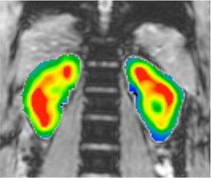
- Lungs (interstitial lung disease, COPD)
- Brain (multiple sclerosis, Alzheimer’s disease)
- Prostate (cancer)
- Breast (cancer)
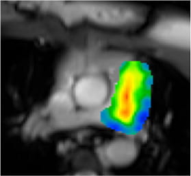
Conventional MRI provides ejection fraction which is currently an important marker for diagnosing many diseases. However, it may not provide a complete picture of cardiac mechanics. Therefore, non-invasive estimate of myocardial stiffness might provide important information regarding mechanical function of the heart. Cardiac MRE has a potential to diagnose a variety of cardiac disease states such as heart failure with preserved ejection fraction (HFPEF), hypertrophic cardiomyopathy (HCM), load independent contractility and myocardial infarction (MI), where stiffness can be an important marker. An example of cardiac MRE performed in a HCM patient is shown below.
Myocardial MRE: Normal (Loop)
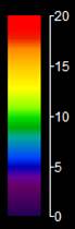
Dynamic Stiffness map (kPa)
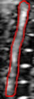
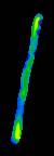
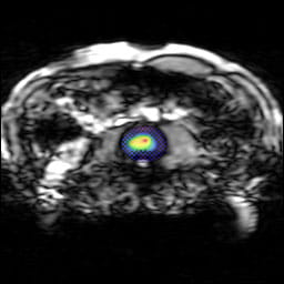
Aortic MRE (Normal Volunteer)
| Magnitude | Wave image | Stiffness map | ||||
 |
 |
 |
 |
Abdominal Aortic Aneurysm
| Magnitude | Wave image | Stiffness map | ||||
 |
 |
 |
|
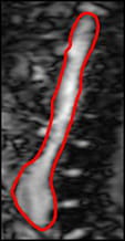
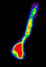
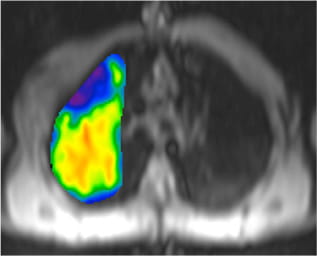
Liver MRE
Stage two fibrosis patient
Magnitude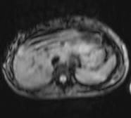
|
Wave image |
Stiffness map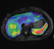 |
||
 |
Wave image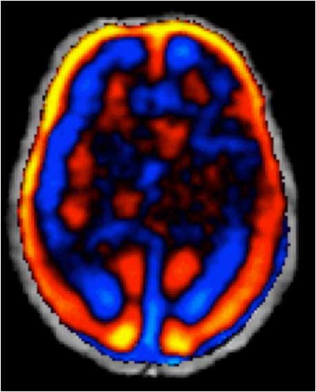
|
Isotropic stiffness (kPa) |
Anisotropic stiffness (kPa)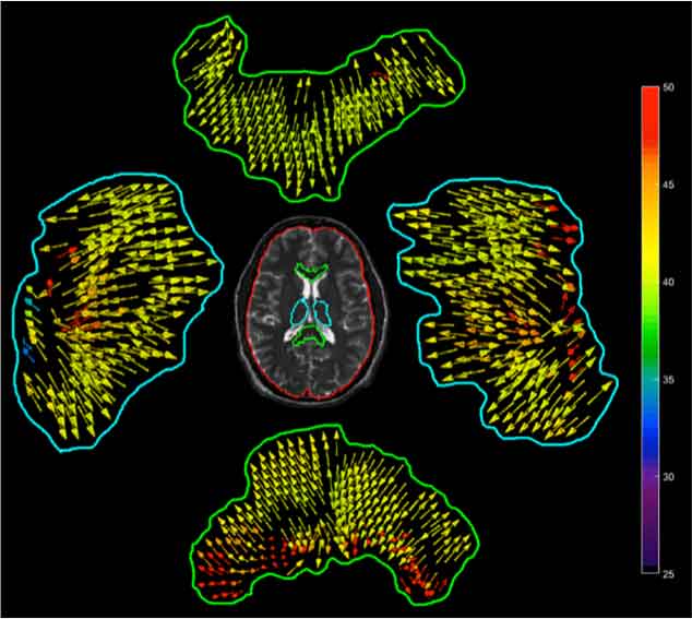 |
Spine |
Kidneys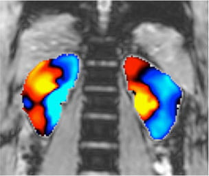 |
Lung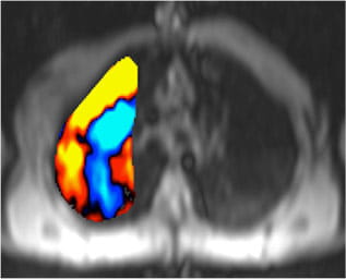 |
Main pulmonary artery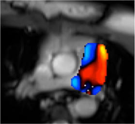 |
|||
 |
|
|
|
|||
 |
 |
|
 |
Program Director