
The Imaging Core Facility is located on the 3rd floor of the Biomedical Research Tower (BRT). These facilities provide high resolution imaging for The Ohio State University researchers using a variety of different instruments.
Our Equipment
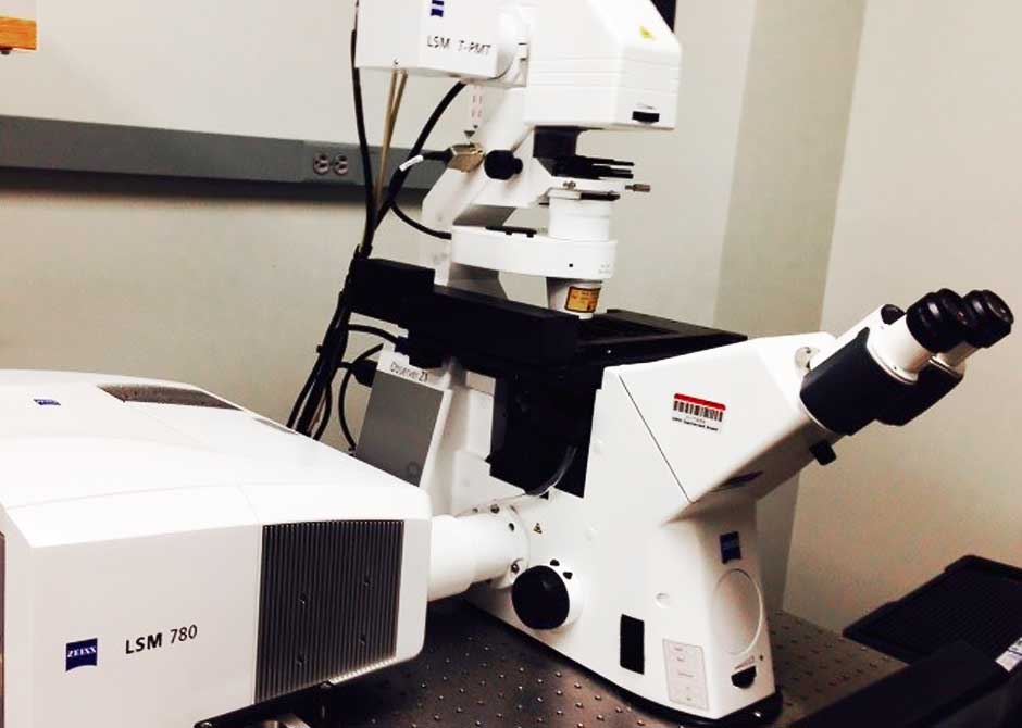
Zeiss LSM 780 Microscope
This microscope supports high-resolution laser scanning confocal microscopic imaging needs. It has a state-of-the-art spectrum detector with high sensitivity, and oil and water immersion high-numerical aperture objectives for examination of live cells and fixed tissue slides. It is equipped with five lasers and transmission light as well.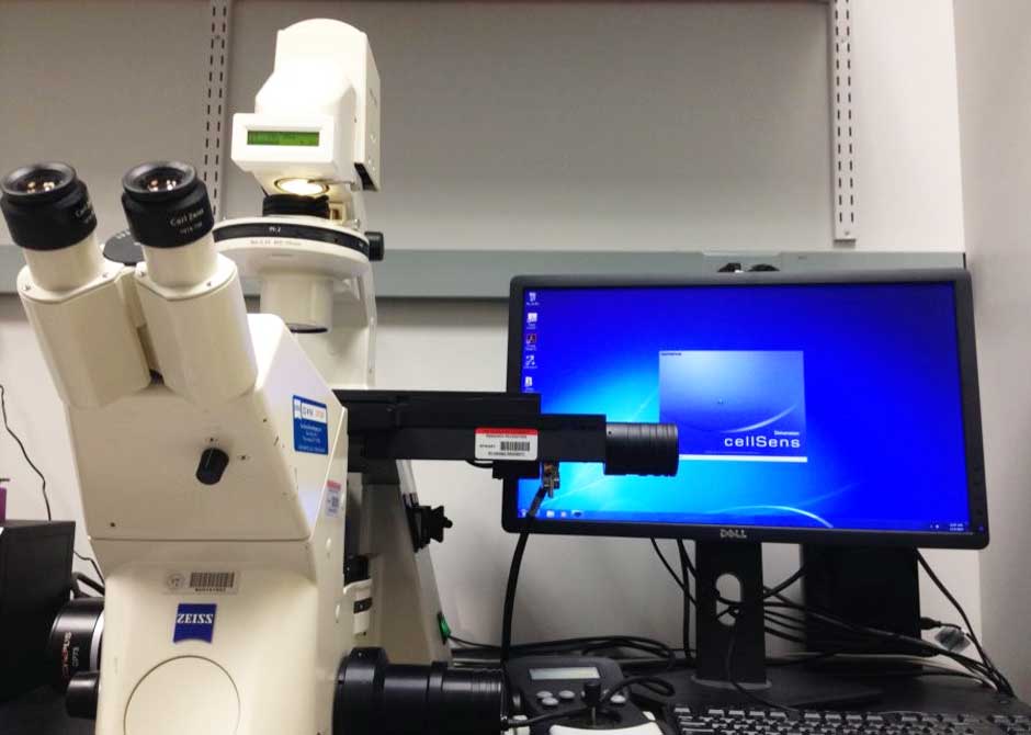
BIO-Scan System for Histology and Pathology Images Storage and Archive
We have a home-made Zeiss Axiovert 200 Bio-Scan system that is built on an inverted Zeiss Axiovert 200 epi-fluorescent microscope. It has 10x, 20x, 40x dry lens and a CCD camera with motorized XY stage. The installed Olympus Cell-Sens Dimension slide reading/archive software allows control of the stage at XY to perform whole slide reading with up to 100x100 montage function.It is thus a very useful tool for both immunohistochemistry and pathology imaging and serves as an archive for storage of large pathological data in a digital format. Through this system, we can share pathological information among research teams.
Our Innovative Imaging Methods
In addition to the conventional Zeiss LSM 780 Microscope, our STORM super-resolution imaging platform has made significant progress by developing several innovative methods of data acquisition, analysis, and image reconstruction. Single-molecule localization microscopy achieves sub-diffraction-limit resolution by localizing a sparse subset of stochastically activated emitters in each frame. Its temporal resolution is limited by the maximal emitter density that can be handled by the image reconstruction algorithms. We developed a new algorithm (MempSTORM) based on two-dimensional spectrum analysis. With the same localization accuracy and recall rate, MempSTORM is 100 times faster than the conventional compressive-sensing-based STORM algorithm.
We also developed a 3D high-density super-resolution imaging platform that allows us to precisely locate the positions of emitters, even when they are significantly overlapped in three-dimensional space.
Moreover, we assembled a new image reconstruction algorithm based on tracklets, short trajectories of the same objects. We improved the localization accuracy by associating the same emitters from multiple frames to form tracklets and by aggregating signals to enhance the signal to noise ratio. Using this method, for the first time, we resolve the transverse tubule structure of the mammalian skeletal muscle.