
 Jun Liu, PhD and her research team are looking at corneal biomechanics in a new way. Funded by a National Institute of Health grant and with software they developed over the past 10 years, the team is using an ultrasonic device, the Ocular Pulse Elastography, to view not only the structure of the cornea, but the function as well.
“We are mapping out the biomechanical responses of the cornea," Liu explains. "We use a high-resolution ultrasound and our algorithms to detect very small corneal deformations in response to the ocular pulse.”
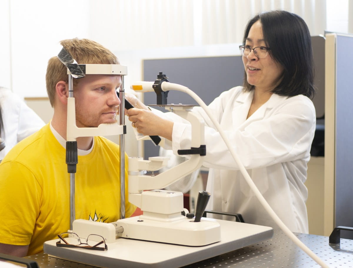
After receiving numbing eye drops, the patient places their chin on the rest and their head is Velcro'd to the top of the device for stability.
The ultrasonic probe, covered first with a cellulose membrane and then standard eye lubricating gel, is lightly touched against the patient's eye. The team is developing new ultrasound gels for easier imaging and obtaining a better ultrasound reading.
"Topography is currently the method for diagnosing keratoconus, but it can only see the shape of the cornea. It doesn't show the mechanical properties; the functional aspects of the cornea," Liu explains.
"Our goal is to provide a functional evaluation. Hopefully this will better capture the early developments in eye problems, for example some people who go through refractive surgery have complications. We hope we can screen people and prevent these complications."
Along with the team’s advanced signal processing algorithms, the ultrasound can capture very minute changes of the cornea—as small as a half-micron change in thickness. The ultrasound aims to measure the pulsation within the eye, but the researchers found that there is a larger pulsation of the whole eye. These extra motions, including building vibrations, can interfere with the measurements.
"If we can analyze the pulsation of the eye, we can get information about the functional aspects of the eye," Liu explains. "The ultrasound is one type of imaging system, and we're doing more than just seeing the structure."
The system is not in-clinic yet, although the team is working hard to reach that goal.
"We're hoping that this software can be a tag-along to a clinical system, so people who already have an ultrasound can use this software with it for their measurements," Liu says.
Liu has been working with Ohio State ophthalmologist Andrew Hendershot, MD to begin measuring patients and obtainingclinical data to better understand diseases like keratoconus and glaucoma.
"Out of all our projects, this is the one that is closest to potential clinical use," Liu says. "We’re very excited about that aspect.”
Research EYElight
“There is something fascinating about how we can see, and what factors can interfere, and what physics, mechanics and optics—all these engineering-based science and technologies—can help improve the healthcare of vision," Dr. Liu says. “My interest has been engineering innovation, but I have always been interested in medicine and healthcare as well.”For Liu, learning and developing new ideas and techniques are another part of why she loves her job.
"There are always new things in the field, in technologies, in questions we ask. There are always new ideas and new possibilities."