
Structural and Functional Assessments (SFA) Core
Director: Andy Fischer, PhD
Co-director: Colleen Cebulla, MD, PhD
Lab manager: Tyler Heisler-Taylor, PhD
ERG: Julie Racine, PhD
Email: vsrcpmanagement@buckeyemail.osu.edu
Please cite P30EY032857 in all publications that make use of the Ohio State University Vision Science Research Core Program (OSU-VSRCP).
“Services were performed at the OSU Vision Sciences Research Core Program under P30EY032857.”
Services include:
- Technician time & Assisted equipment use
- Animal handling, husbandry, or other expertise
- Equipment or procedure training
- Contract work with the Center for Design and Manufacturing Excellence (CDME)
-
3D Printing, Electronics, Machining, Material Testing, Metal Casting, Polymer Processing, Precision Cutting, Robotics, Welding, and More!
-
Please reach out or schedule a consultation through eRAMP!
Equipment includes:
The Celeris ERG System is able to record light- and dark-adapted ERG measurements on a range of species from mice to rabbits. It uses a novel alternating electrode-stimulator to quickly acquire ERG recordings. Equipment for mice, rats, chickens, and rabbits available.
Owner: Dr. Matthew Reilly
Location: Wiseman Hall N212
The E3 ERG Systems is able to record light- and dark- adapted full field ERG measurements on many species from mouse to human utilizing a ganzfeld dome and handheld light stimulator.
Owner: Dr. Thomas Mendel
Location: Wiseman Hall N212 & PRC 4170A
The iCare Tonovet Plus is a rebound tonometer used to measure the IOP. This model is calibrated for larger species (rabbit to horse), the extremely light contact of the probe is so gentle that even mice can be measured without any noticeable reflex.
Owner: Dr. Colleen Cebulla
Location: Wiseman Hall N212
 Two HM550 cryostats and a new Dakewe 2650 cryostat are available for core users for tissue cryosectioning. Slides, OCT mounting solution, and cryostat blades can be provided but those using the cryostat are encouraged to use their own to keep supplies available to all.
Owner: Dr. Colleen Cebulla & Dr. Andrew Fischer
Location: Wiseman Hall N212, Graves Hall 3041, & PRC 4170
 The Leica Envisu SD-OCT is a spectral domain optical coherence tomography (SD-OCT) system capable of acquiring ‘optical biopsies’, cross sections of the retina in a 3D volume in real time on live subjects. We have mouse and rat/chicken mounts, adjustments can be made for other species. We also have mouse, rat/chicken, and a corneal/in vitro telecentric bore lens.
Manager Assistance Required
Owner: Dr. Colleen Cebulla
Location: Wiseman Hall N212
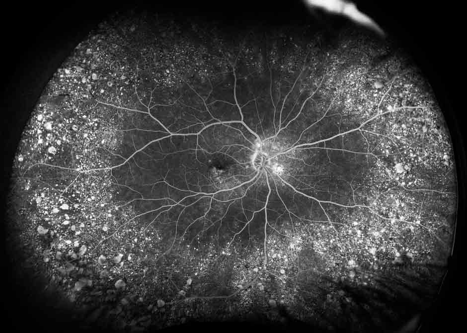
 The Micron IV retinal imaging microscope allows bright-field, fluorescien and Evan's blue angiography, and fluorescent imaging of common report molecules. The image-guided laser delivery module allows real-time targeting for photocoagulation damage models. The MICRON Image-Guided OCT2 system in eludes live, real-time fundus display with precise positioning to capture OCT scans. Training to be provided on request for equipment and software.
 Use Requires Dr. Kerur's Approval
Owner: Dr. Nagaraj Kerur
Location: Pelotonia Research Center 4130U
The Nikon AXR confocal microscope point scan microscope with the NIS-Elements Advanced Research package allows high-quality imaging. The Advanced Research package includes state-of-the-art AI algorithmic analysis. We additionally have a remote access station available for approved users.
Department of Ophthalmology & Visual Sciences faculty & staff are provided unlimited free use of Nikon AXR Confocal.
Owner: Dr. Sayoko Moroi
Location: Wiseman Hall 1007

The Crest Optics X-Light V3 on a Nikon Ti2 microscope is a spinning disk confocal system. The high speed imaging capabilities make this an ideal system for live-cell imaging experiments. Both confocal systems utilize the NIS-Elements platform allowing cross-compatibility between workstations.
Department of Ophthalmology & Visual Sciences faculty & staff are provided unlimited free use of the X-Light V3.
Owner: Dr. Sayoko Moroi
Location: PRC 4130K
The Operating Microscope allows users a magnified view for precise operations. There are 2 view ports and a fine focus camera for on-screen view. Additionally, a foot pedal allows for easy manipulation of the magnification and focus level.
Owner: Dr. Colleen Cebulla
Location: Wiseman Hall N212
 The Ivesta 3 stereo microscope provides a high working distance operating area for high precision tasks. See website for additional product information.
Owner: Dr. Nagaraj Kerur
Location: PRC 4130
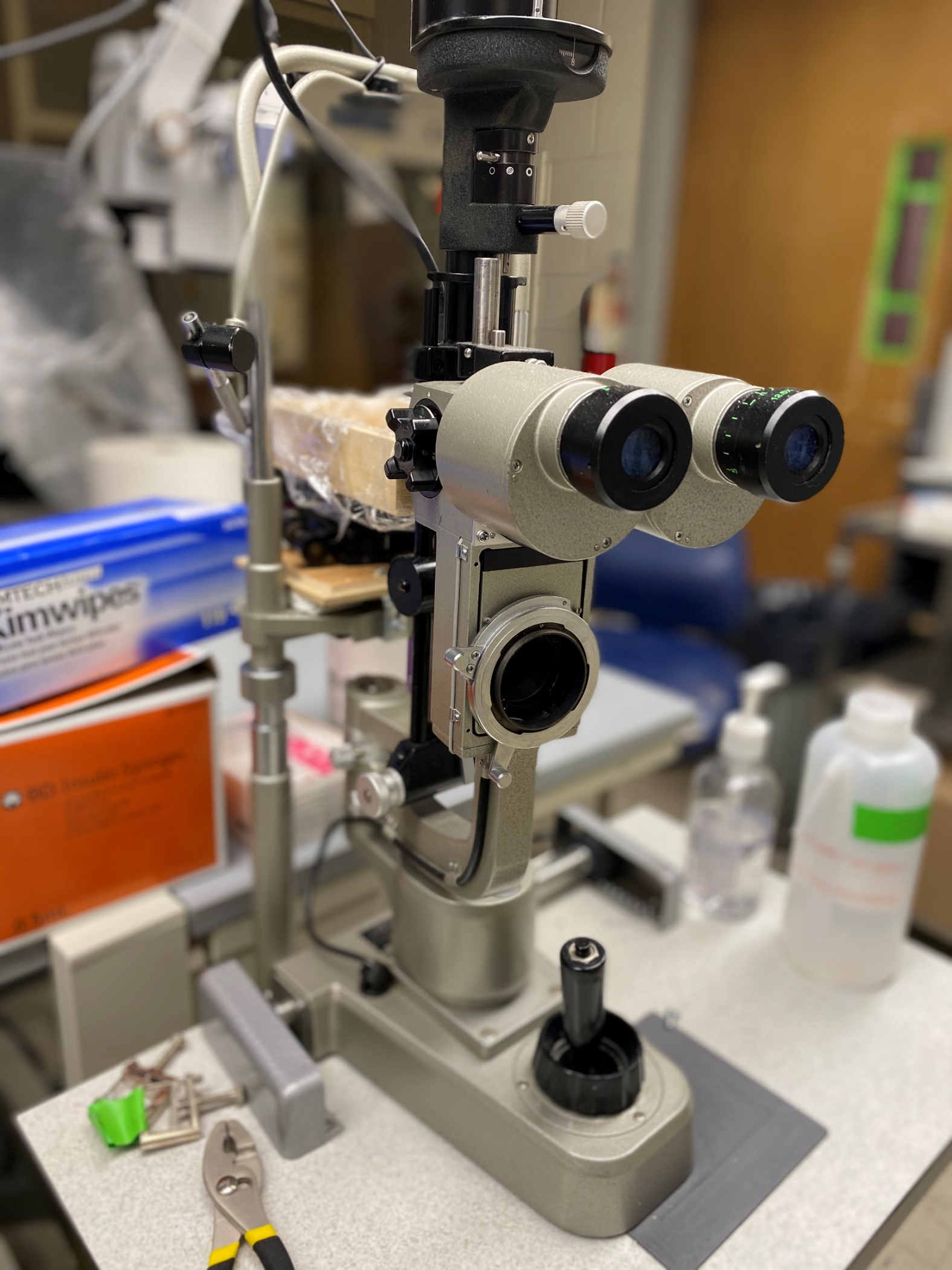
A slit lamp is available for topical and anterior segment observation.
Owner: Dr. Colleen Cebulla
Location: Wiseman Hall N212
 Striatech's OptoDrum automatically determines the visual acuity and contrast sensitivity of mice and rats.
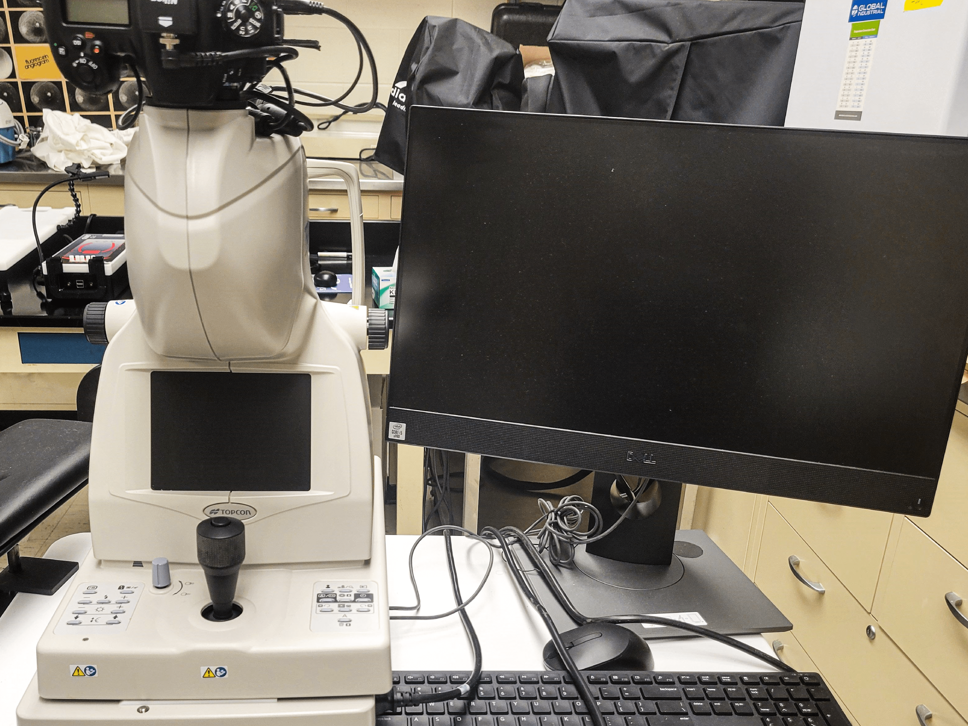
The Topcon TRC-NW8F fundus camera provides color, red-free, and fluorescent angiography for in vivo applications.
Owner: Dr. Thomas Mendel
Location: Wiseman Hall 1007
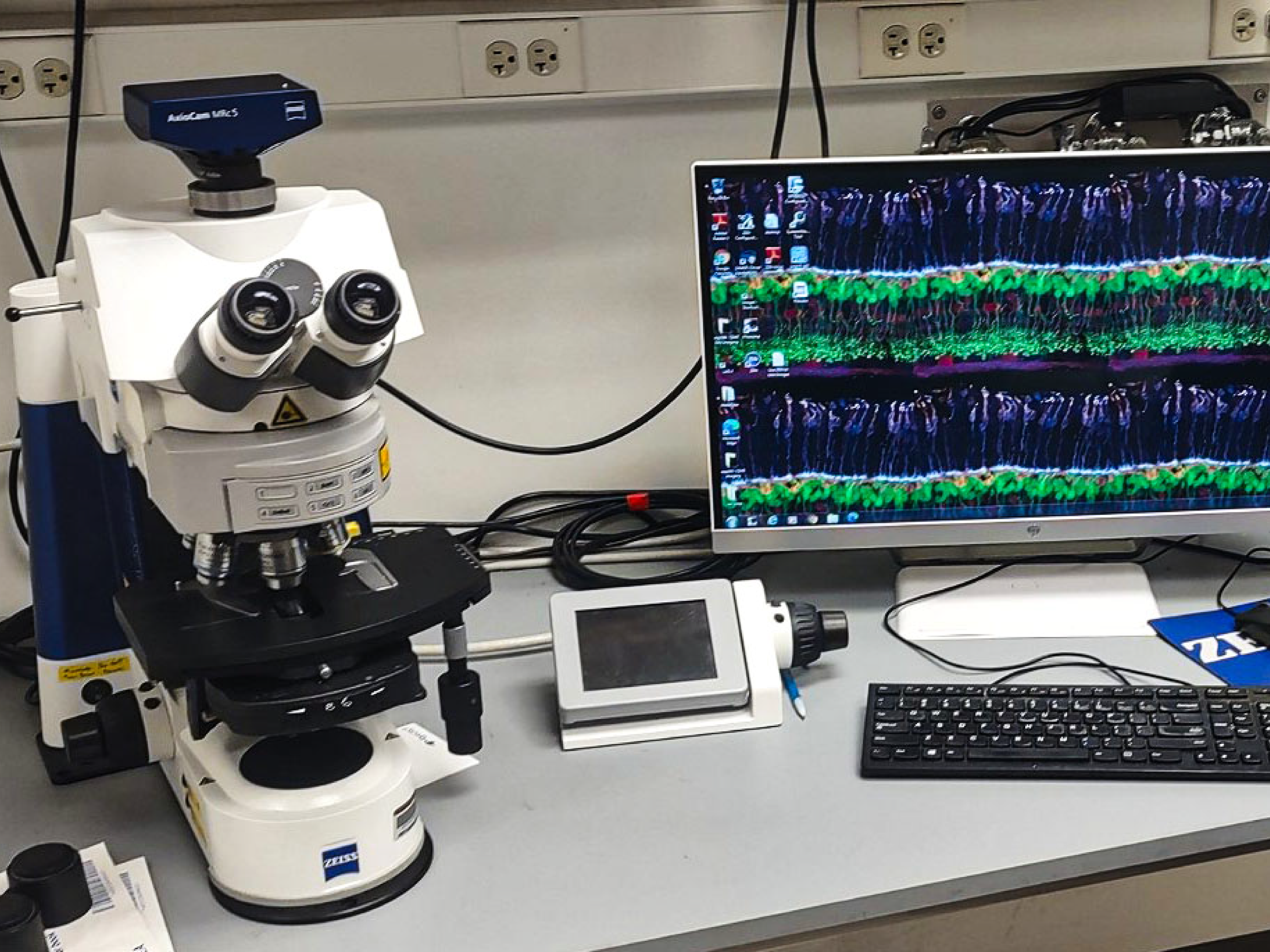
The Axio Imager microscope is an upright fluorescent microscope for imaging stained samples.
Owner: Dr. Andrew Fischer
Location: Graves Hall 3041
Expanded equipment access and data gathering on structural and functional analysis
Core A focuses on physiological and structural data gathered from the cellular level (in vitro) to animal models (in vivo) of eye diseases. The work often requires expensive equipment and technical expertise that individual researchers can’t afford but institutions can, such as:
- Angiography
- Electrophysiology
- Fundus photography
- Photocoagulation laser systems
- State-of-the-art microscopes
- Optical coherence tomography
- Tonometry
The Core A team, directed by DOVS faculty Colleen Cebulla, MD, PhD, and Department of Neuroscience faculty Andy Fischer, PhD, will also provide consulting expertise to scientists and labs that need additional analysis or help with developing specific research methods. Such technical support can also assist funded scientists in completing additional studies within their funded projects
For example, when studying macular degeneration, glaucoma or inherited retinal dystrophy, a researcher may study one aspect of a condition, such as histology. The P30 cores offer the ability for the same scientist to expand their work by taking in vivo pictures over time and some electrophysiologic functions of the diseased tissues or eyes.
“This P30 grant will enhance research of funded investigators and also help faculty without grants who need critical pilot data in order to write a strong new grant proposal." - Dr. Moroi.